Explore
 Validate
 Learn
Learn Western blot
 Immunocytochemistry
Antibody data
- Antibody Data
- Antigen structure
- References [0]
- Comments [0]
- Validations
- Immunocytochemistry [2]
Submit
Validation data
Reference
Comment
Report error
- Product number
- PA5-95950 - Provider product page

- Provider
- Invitrogen Antibodies
- Product name
- Syndecan 4 Polyclonal Antibody
- Antibody type
- Polyclonal
- Antigen
- Recombinant full-length protein
- Description
- Immunogen sequence: ESIRETEVID PQDLLEGRYF SGALPDDEDV VGPGQESDDF ELSGSGDLDD LEDSMIGPEV VHPLVPLDNH IPERAGSGSQ VPTEPKKLEE NEVIPKRISP VEESEDVSNK VSMSSTVQGS NIFERTE; Positive Samples: Mouse kidney; Cellular Location: Membrane, Single-pass type I membrane protein
- Reactivity
- Human
- Host
- Rabbit
- Isotype
- IgG
- Vial size
- 100 µL
- Concentration
- 2.02 mg/mL
- Storage
- -20° C, Avoid Freeze/Thaw Cycles
No comments: Submit comment
Supportive validation
- Submitted by
- Invitrogen Antibodies (provider)
- Main image
- Experimental details
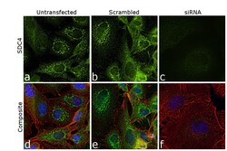
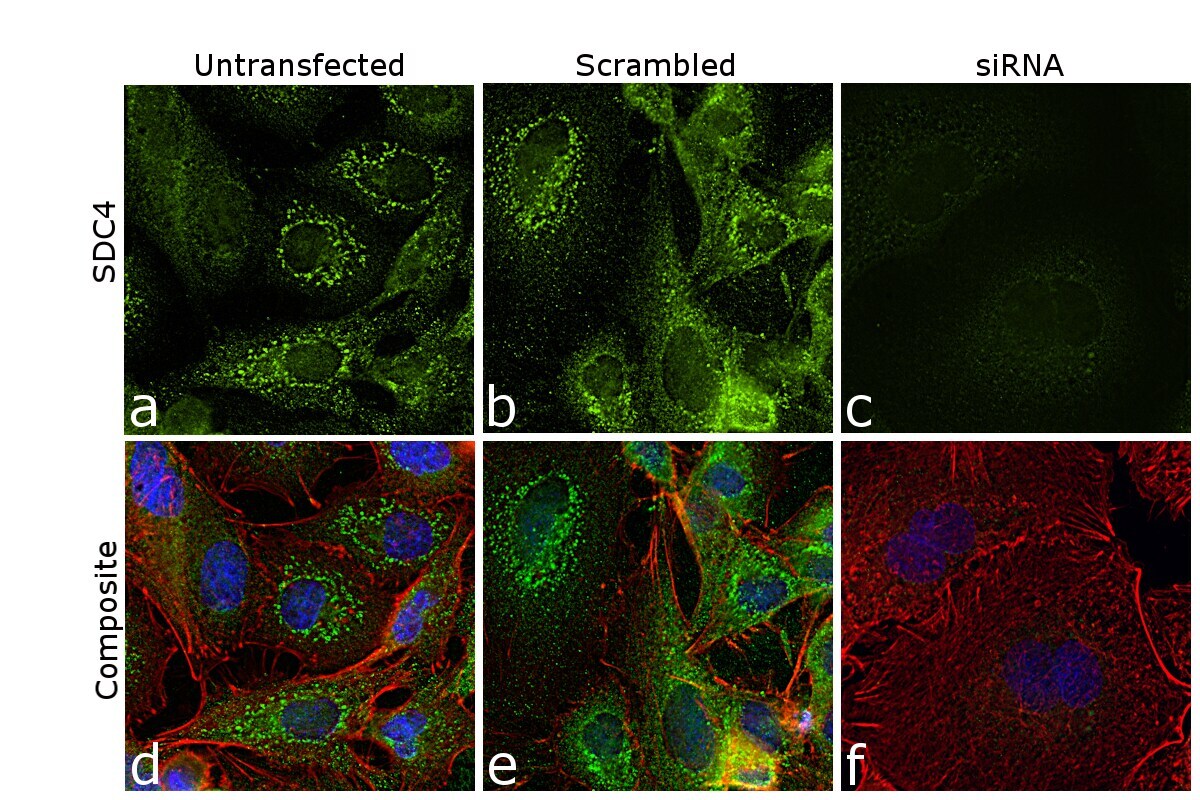
- Knockdown of SDC4 was achieved by transfecting A549 cells with SDC4-specific siRNA (Silencer® select Product # S12638, S12640). Immunofluorescence analysis was performed on untransfected A549 cells (panels a,d), transfected with non-specific scrambled siRNA (panels b,e), and transfected with SDC4-specific siRNA (panels c,f). Cells were fixed, permeabilized, and labeled with Syndecan 4 Polyclonal Antibody (Product # PA5-95950, 1:100 dilution) followed by Donkey anti-Rabbit IgG (H+L) Highly Cross-Adsorbed Secondary Antibody, Alexa Fluor Plus 488 (Product # A32790, 1:2000 dilution). Nuclei (blue) were stained using ProLong™ Diamond Antifade Mountant with DAPI (Product # P36962), and Rhodamine Phalloidin (Product # R415, 1:300) was used for cytoskeletal F-actin (Red) staining. Reduction of specific signal was observed upon siRNA mediated knockdown (panel c,f) confirming specificity of the antibody to SDC4 (Green). The Images were captured at 60X magnification.
- Submitted by
- Invitrogen Antibodies (provider)
- Main image
- Experimental details
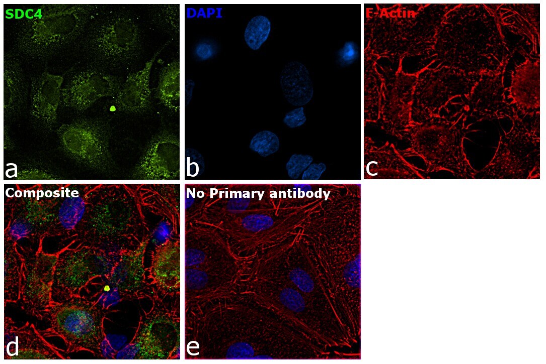
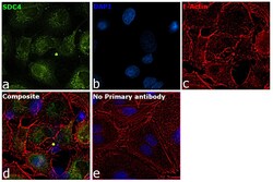
- Immunofluorescence analysis of SDC4 was performed using 70% confluent log phase A549 cells. The cells were fixed with 4% paraformaldehyde for 10 minutes, permeabilized with 0.1% Triton™ X-100 for 15 minutes, and blocked with 2% BSA for 45 minutes at room temperature. The cells were labeled with Syndecan 4 Polyclonal Antibody (Product # PA5-95950) at 1:100 dilution in 0.1% BSA, incubated at 4 degrees Celsius overnight, and then labeled with Donkey anti-Rabbit IgG (H+L) Highly Cross-Adsorbed Secondary Antibody, Alexa Fluor Plus 488 (Product # A32790, 1:2000 dilution), for 45 minutes at room temperature (Panel a: Green). Nuclei (Panel b: Blue) were stained with ProLong™ Diamond Antifade Mountant with DAPI (Product # P36962). F-actin (Panel c: Red) was stained with Rhodamine Phalloidin (Product # R415, 1:300). Panel d represents the merged image showing Golgi localization. Panel e represents control cells with no primary antibody to assess the background. The images were captured at 60X magnification.