Explore
 Validate
 Learn
Learn Western blot
 Immunohistochemistry
Antibody data
- Antibody Data
- Antigen structure
- References [3]
- Comments [0]
- Validations
- Immunohistochemistry [1]
Submit
Validation data
Reference
Comment
Report error
- Product number
- AMAb90667 - Provider product page

- Provider
- Atlas Antibodies
- Proper citation
- Atlas Antibodies Cat#AMAb90667, RRID:AB_2665627
- Product name
- Anti-PODXL
- Antibody type
- Monoclonal
- Description
- Monoclonal Antibody against Human PODXL, Clone ID: CL0308, Gene description: podocalyxin-like, Alternative Gene Names: Gp200, PC, PCLP, Validated applications: WB, IHC, Uniprot ID: O00592, Storage: Store at +4°C for short term storage. Long time storage is recommended at -20°C.
- Reactivity
- Human
- Host
- Mouse
- Conjugate
- Unconjugated
- Isotype
- IgG
- Antibody clone number
- CL0308
- Vial size
- 100 µl
- Concentration
- 1.0 mg/ml
- Storage
- Store at +4°C for short term storage. Long time storage is recommended at -20°C.
- Handling
- The antibody solution should be gently mixed before use.
Submitted references Podocalyxin-like and RNA-binding motif protein 3 are prognostic biomarkers in urothelial bladder cancer: a validatory study.
Membranous expression of podocalyxin-like protein is an independent factor of poor prognosis in urothelial bladder cancer
Membranous expression of podocalyxin-like protein is an independent factor of poor prognosis in urothelial bladder cancer.
Boman K, Andersson G, Wennersten C, Nodin B, Ahlgren G, Jirström K
Biomarker research 2017;5:10
Biomarker research 2017;5:10
Membranous expression of podocalyxin-like protein is an independent factor of poor prognosis in urothelial bladder cancer
Boman K, Larsson A, Segersten U, Kuteeva E, Johannesson H, Nodin B, Eberhard J, Uhlén M, Malmström P, Jirström K
British Journal of Cancer 2013 May;108(11):2321-2328
British Journal of Cancer 2013 May;108(11):2321-2328
Membranous expression of podocalyxin-like protein is an independent factor of poor prognosis in urothelial bladder cancer.
Boman K, Larsson AH, Segersten U, Kuteeva E, Johannesson H, Nodin B, Eberhard J, Uhlén M, Malmström PU, Jirström K
British journal of cancer 2013 Jun 11;108(11):2321-8
British journal of cancer 2013 Jun 11;108(11):2321-8
No comments: Submit comment
Supportive validation
- Submitted by
- Atlas Antibodies (provider)
- Enhanced method
- Orthogonal validation
- Main image
- Experimental details
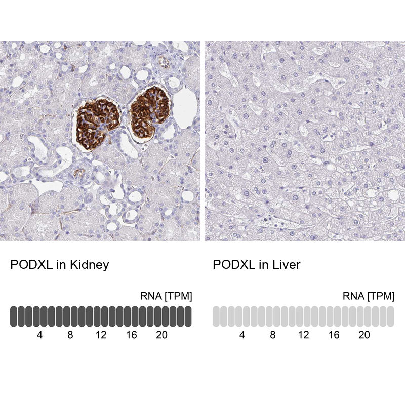
- Immunohistochemistry analysis in human kidney and liver tissues using AMAb90667 antibody. Corresponding PODXL RNA-seq data are presented for the same tissues.
- Sample type
- Human
- Protocol
- Protocol