Explore
 Validate
 Learn
Learn Western blot
 Immunocytochemistry
 Immunoprecipitation
Antibody data
- Antibody Data
- Antigen structure
- References [5]
- Comments [0]
- Validations
- Immunocytochemistry [3]
- Immunohistochemistry [3]
- Other assay [1]
Submit
Validation data
Reference
Comment
Report error
- Product number
- PA1-042 - Provider product page

- Provider
- Invitrogen Antibodies
- Product name
- ADORA2A Polyclonal Antibody
- Antibody type
- Polyclonal
- Antigen
- Synthetic peptide
- Description
- PA1-042 detects adenosine receptor A2a (A2aAR) in human, rat, mouse and canine tissues. This antibody does not detect other AR subtypes. PA1-042 has been successfully used in Western blot, ICC/IF, immunohistochemistry and immunoprecipitation procedures. By Western blot, this antibody detects an ~45 kDa protein from canine striatum representing A2aAR. Immunohistochemical staining of A2aAR in human hippocampus with PA1-042 yields a pattern consistent with plasma membrane staining. The PA1-042 immunogen is a synthetic peptide corresponding to residues E(373) S H G D M G L P D V E L L S H E L K(391) of canine A2aAR.
- Reactivity
- Human, Mouse, Rat, Canine
- Host
- Rabbit
- Isotype
- IgG
- Vial size
- 100 μL
- Concentration
- 0.69 mg/mL
- Storage
- -20°C, Avoid Freeze/Thaw Cycles
Submitted references A venous-specific purinergic signaling cascade initiated by Pannexin 1 regulates TNFα-induced increases in endothelial permeability.
On the Role of Adenosine A2A Receptor Gene Transcriptional Regulation in Parkinson's Disease.
Abnormal calcium handling in atrial fibrillation is linked to up-regulation of adenosine A2A receptors.
Adenosine A 2B receptors modulate cAMP levels and induce CREB but not ERK1/2 and p38 phosphorylation in rat skeletal muscle cells.
Stimulation of A(2A) adenosine receptor phosphorylation by protein kinase C activation: evidence for regulation by multiple protein kinase C isoforms.
Maier-Begandt D, Comstra HS, Molina SA, Krüger N, Ruddiman CA, Chen YL, Chen X, Biwer LA, Johnstone SR, Lohman AW, Good ME, DeLalio LJ, Hong K, Bacon HM, Yan Z, Sonkusare SK, Koval M, Isakson BE
Science signaling 2021 Mar 2;14(672)
Science signaling 2021 Mar 2;14(672)
On the Role of Adenosine A2A Receptor Gene Transcriptional Regulation in Parkinson's Disease.
Falconi A, Bonito-Oliva A, Di Bartolomeo M, Massimini M, Fattapposta F, Locuratolo N, Dainese E, Pascale E, Fisone G, D'Addario C
Frontiers in neuroscience 2019;13:683
Frontiers in neuroscience 2019;13:683
Abnormal calcium handling in atrial fibrillation is linked to up-regulation of adenosine A2A receptors.
Llach A, Molina CE, Prat-Vidal C, Fernandes J, Casadó V, Ciruela F, Lluís C, Franco R, Cinca J, Hove-Madsen L
European heart journal 2011 Mar;32(6):721-9
European heart journal 2011 Mar;32(6):721-9
Adenosine A 2B receptors modulate cAMP levels and induce CREB but not ERK1/2 and p38 phosphorylation in rat skeletal muscle cells.
Lynge J, Schulte G, Nordsborg N, Fredholm BB, Hellsten Y
Biochemical and biophysical research communications 2003 Jul 18;307(1):180-7
Biochemical and biophysical research communications 2003 Jul 18;307(1):180-7
Stimulation of A(2A) adenosine receptor phosphorylation by protein kinase C activation: evidence for regulation by multiple protein kinase C isoforms.
Palmer TM, Stiles GL
Biochemistry 1999 Nov 9;38(45):14833-42
Biochemistry 1999 Nov 9;38(45):14833-42
No comments: Submit comment
Supportive validation
- Submitted by
- Invitrogen Antibodies (provider)
- Main image

- Experimental details
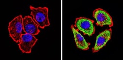
- Immunofluorescent analysis of Adenosine Receptor A2a (green) showing staining in the cytoplasm of U251 cells (right) compared to a negative control without primary antibody (left). Formalin-fixed cells were permeabilized with 0.1% Triton X-100 in TBS for 5-10 minutes and blocked with 3% BSA-PBS for 30 minutes at room temperature. Cells were probed with an Adenosine Receptor A2a polyclonal antibody (Product # PA1-042) in 3% BSA-PBS at a dilution of 1:20 and incubated overnight at 4 ºC in a humidified chamber. Cells were washed with PBST and incubated with a DyLight-conjugated secondary antibody in PBS at room temperature in the dark. F-actin (red) was stained with a fluorescent red phalloidin and nuclei (blue) were stained with Hoechst or DAPI. Images were taken at a magnification of 60x.

- Submitted by
- Invitrogen Antibodies (provider)
- Main image

- Experimental details
- Immunofluorescent analysis of Adenosine Receptor A2a (green) showing staining in the cytoplasm of U251 cells (right) compared to a negative control without primary antibody (left). Formalin-fixed cells were permeabilized with 0.1% Triton X-100 in TBS for 5-10 minutes and blocked with 3% BSA-PBS for 30 minutes at room temperature. Cells were probed with an Adenosine Receptor A2a polyclonal antibody (Product # PA1-042) in 3% BSA-PBS at a dilution of 1:20 and incubated overnight at 4 ºC in a humidified chamber. Cells were washed with PBST and incubated with a DyLight-conjugated secondary antibody in PBS at room temperature in the dark. F-actin (red) was stained with a fluorescent red phalloidin and nuclei (blue) were stained with Hoechst or DAPI. Images were taken at a magnification of 60x.

- Submitted by
- Invitrogen Antibodies (provider)
- Main image
- Experimental details
- Immunofluorescent analysis of Adenosine Receptor A2a (green) showing staining in the cytoplasm of U251 cells (right) compared to a negative control without primary antibody (left). Formalin-fixed cells were permeabilized with 0.1% Triton X-100 in TBS for 5-10 minutes and blocked with 3% BSA-PBS for 30 minutes at room temperature. Cells were probed with an Adenosine Receptor A2a polyclonal antibody (Product # PA1-042) in 3% BSA-PBS at a dilution of 1:20 and incubated overnight at 4 ºC in a humidified chamber. Cells were washed with PBST and incubated with a DyLight-conjugated secondary antibody in PBS at room temperature in the dark. F-actin (red) was stained with a fluorescent red phalloidin and nuclei (blue) were stained with Hoechst or DAPI. Images were taken at a magnification of 60x.

Supportive validation
- Submitted by
- Invitrogen Antibodies (provider)
- Main image

- Experimental details
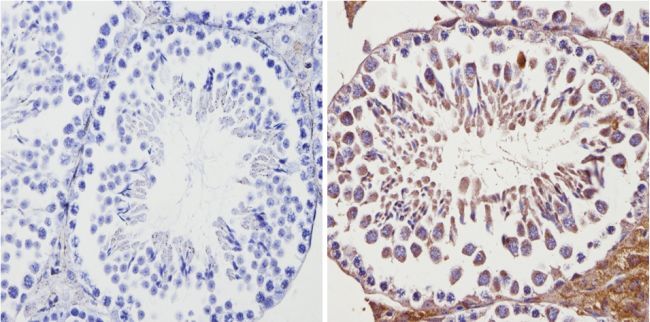
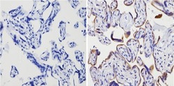
- Immunohistochemistry analysis of Adenosine Receptor A2a showing positive staining in the cytoplasm and membrane of paraffin-treated Mouse testis tissue (right) compared with a negative control in the absence of primary antibody (left). To expose target proteins, antigen retrieval method was performed using 10mM sodium citrate (pH 6.0) microwaved for 8-15 min. Following antigen retrieval, tissues were blocked in 3% H2O2-methanol for 15 min at room temperature, washed with ddH2O and PBS, and then probed with a Adenosine Receptor A2a polyclonal antibody (Product # PA1-042) diluted by 3% BSA-PBS at a dilution of 1:20 overnight at 4°C in a humidified chamber. Tissues were washed extensively PBST and detection was performed using an HRP-conjugated secondary antibody followed by colorimetric detection using a DAB kit. Tissues were counterstained with hematoxylin and dehydrated with ethanol and xylene to prep for mounting.
- Submitted by
- Invitrogen Antibodies (provider)
- Main image
- Experimental details
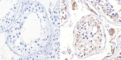
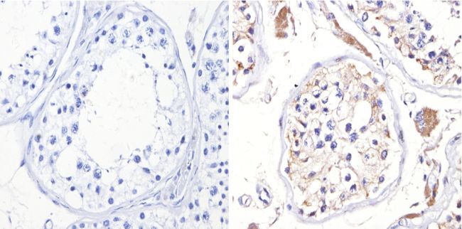
- Immunohistochemistry analysis of Adenosine Receptor A2a showing positive staining in the cytoplasm and membrane of paraffin-treated Human testis tissue (right) compared with a negative control in the absence of primary antibody (left). To expose target proteins, antigen retrieval method was performed using 10mM sodium citrate (pH 6.0) microwaved for 8-15 min. Following antigen retrieval, tissues were blocked in 3% H2O2-methanol for 15 min at room temperature, washed with ddH2O and PBS, and then probed with a Adenosine Receptor A2a polyclonal antibody (Product # PA1-042) diluted by 3% BSA-PBS at a dilution of 1:200 overnight at 4°C in a humidified chamber. Tissues were washed extensively PBST and detection was performed using an HRP-conjugated secondary antibody followed by colorimetric detection using a DAB kit. Tissues were counterstained with hematoxylin and dehydrated with ethanol and xylene to prep for mounting.
- Submitted by
- Invitrogen Antibodies (provider)
- Main image
- Experimental details
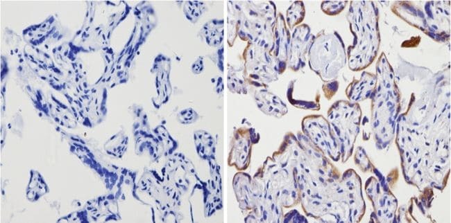
- Immunohistochemistry analysis of Adenosine Receptor A2a showing positive staining in the cytoplasm and membrane of paraffin-treated Human placenta tissue (right) compared with a negative control in the absence of primary antibody (left). To expose target proteins, antigen retrieval method was performed using 10mM sodium citrate (pH 6.0) microwaved for 8-15 min. Following antigen retrieval, tissues were blocked in 3% H2O2-methanol for 15 min at room temperature, washed with ddH2O and PBS, and then probed with a Adenosine Receptor A2a polyclonal antibody (Product # PA1-042) diluted by 3% BSA-PBS at a dilution of 1:100 overnight at 4°C in a humidified chamber. Tissues were washed extensively PBST and detection was performed using an HRP-conjugated secondary antibody followed by colorimetric detection using a DAB kit. Tissues were counterstained with hematoxylin and dehydrated with ethanol and xylene to prep for mounting.
Supportive validation
- Submitted by
- Invitrogen Antibodies (provider)
- Main image
- Experimental details
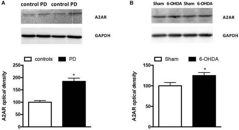
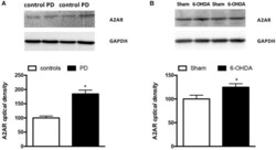
- FIGURE 4 Analysis of A2AR protein levels in PBMCs from (A) PD patients ( n = 5) and controls ( n = 4) and (B) Sham ( n = 12) and 6-OHDA ( n = 10) mice striata. Representative immunoblots of PBMCs and striata lysates reacted with specific anti-A2AR or anti-GAPDH antibodies are shown above the bars for both mice and humans. Values, expressed as means +- standard error of the mean (SEM), were normalized by GAPDH and taking the control and sham groups as 100, respectively for (A,B) . * p < 0.05 vs. respective control groups.