 Explore
 Validate
 Learn
Learn Western blot
 ELISA
ELISAAntibody data
- Antibody Data
- Antigen structure
- References [1]
- Comments [0]
- Validations
- Western blot [6]
- Immunohistochemistry [2]
Submit
Validation data
Reference
Comment
Report error
- Product number
- NBP2-25092 - Provider product page

- Provider
- Novus Biologicals
- Product name
- Rabbit Polyclonal TRAILR1/TNFRSF10A Antibody
- Antibody type
- Polyclonal
- Description
- Peptide affinity purified.
- Reactivity
- Human
- Host
- Rabbit
- Isotype
- IgG
- Vial size
- 0.1 mg
- Concentration
- 1 mg/ml
- Storage
- Store at 4C short term. Aliquot and store at -20C long term. Avoid freeze-thaw cycles.
Submitted references Synergistic antitumor activity of TRAIL combined with chemotherapeutic agents in A549 cell lines in vitro and in vivo.
Fan QL, Zou WY, Song LH, Wei W
Cancer chemotherapy and pharmacology 2005 Feb;55(2):189-96
Cancer chemotherapy and pharmacology 2005 Feb;55(2):189-96
No comments: Submit comment
Supportive validation
- Submitted by
- Novus Biologicals (provider)
- Main image

- Experimental details
- Western Blot: TRAILR1/TNFRSF10A Antibody [NBP2-25092] - Analysis of TRAIL R1/TNFRSF10A in HeLa total cell lysate with TRAIL R1/TNFRSF10A antibody at 0.5 ug/mL.

- Submitted by
- Novus Biologicals (provider)
- Main image

- Experimental details
- Western Blot: TRAILR1/TNFRSF10A Antibody [NBP2-25092] - Analysis of 50 ug of total cell lysate from HeLa cells with anti-DR4 (CT) at 1:500 dilution.

- Submitted by
- Novus Biologicals (provider)
- Main image

- Experimental details
- Western Blot: TRAILR1/TNFRSF10A Antibody [NBP2-25092] - Analysis of DR4 in (A) HepG2 and (B) Jurkat cell lysate with DR4 antibody at 1 ug/ml.

- Submitted by
- Novus Biologicals (provider)
- Main image

- Experimental details
- Western Blot: TRAILR1/TNFRSF10A Antibody [NBP2-25092] - Independent Antibody Validation (IAV) via Protein Expression Profile in Cell Lines Loading: 15 ug of lysates per lane. Antibodies: DR4 NBP2-25092 (1 ug/mL), DR4 NBP1-76473 ( 4 ug/mL), and beta-actin (1 ug/mL), 1h incubation at RT in 5% NFDM/TBST. Secondary: Goat anti-rabbit IgG HRP conjugate.

- Submitted by
- Novus Biologicals (provider)
- Main image

- Experimental details
- Western Blot: TRAILR1/TNFRSF10A Antibody [NBP2-25092] - Western Blot Validation in Cell Lines. Loading: 15 ug of cell lysates per lane. Antibodies: DR4 NBP2-25092 (1 ug/mL), 1h incubation at RT in 5% NFDM/TBST. Secondary: Goat anti-rabbit IgG HRP conjugate at 1:10000 dilution.

- Submitted by
- Novus Biologicals (provider)
- Main image

- Experimental details
- Western Blot: TRAILR1/TNFRSF10A Antibody [NBP2-25092] - KO Validation in HeLa Cells. Loading: 10 ug of HeLa WT cell lysates or TRAILR1/TNFRSF10A KO cell lysates. Antibodies: TRAILR1/TNFRSF10A [NBP2-25092] (1 ug/ml) and beta-actin [NBP1-76692] (1 ug/ml), 1h incubation at RT in 5% NFDM/TBST. Secondary: Goat anti-rabbit IgG HRP conjugate at 1:10000 dilution.

Supportive validation
- Submitted by
- Novus Biologicals (provider)
- Main image
- Experimental details
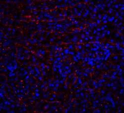
- Immunohistochemistry: TRAILR1/TNFRSF10A Antibody [NBP2-25092] - Immunofluorescence Validation of DR4. Immunofluorescent analysis of 4% paraformaldehyde-fixed human spleen tissue labeling DR4 with NBP2-25092 at 20 ug/mL, followed by goat anti-rabbit IgG secondary antibody (red) and DAPI staining (blue). Image showing membrane staining on human spleen cells.

- Submitted by
- Novus Biologicals (provider)
- Main image
- Experimental details
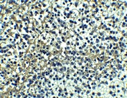
- Immunohistochemistry: TRAILR1/TNFRSF10A Antibody [NBP2-25092] - Staining of human spleen tissue with antibody at 5 ug/mL.
