Explore
 Validate
 Learn
LearnMA1-25373
antibody from Invitrogen Antibodies
Targeting: TNFRSF10A
Apo2, CD261, DR4, TRAILR-1, TRAILR1
 Western blot
 Flow cytometry
Antibody data
- Antibody Data
- Antigen structure
- References [0]
- Comments [0]
- Validations
- Western blot [5]
Submit
Validation data
Reference
Comment
Report error
- Product number
- MA1-25373 - Provider product page

- Provider
- Invitrogen Antibodies
- Product name
- DR4 Monoclonal Antibody (32A1380)
- Antibody type
- Monoclonal
- Antigen
- Synthetic peptide
- Description
- MA1-25373 detects TRAIL Receptor 1 from human samples.
- Reactivity
- Human
- Host
- Mouse
- Isotype
- IgG
- Antibody clone number
- 32A1380
- Vial size
- 50 µg
- Concentration
- 0.5 mg/mL
- Storage
- Store at 4°C short term. For long term storage, store at -20°C, avoiding freeze/thaw cycles.
No comments: Submit comment
Supportive validation
- Submitted by
- Invitrogen Antibodies (provider)
- Main image

- Experimental details
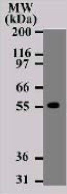
- Western blot of TRAIL-R1/DR4 in Daudi cell lysate using a TRAIL-R1/DR4 monoclonal antibody (Product # MA1-25373) at a dilution of 2 µg/mL followed by detection using a goat anti-mouse Ig HRP secondary antibody and PicoTect ECL substrate solution.

- Submitted by
- Invitrogen Antibodies (provider)
- Main image

- Experimental details
- Western Blot analysis of DR4 was performed by loading Daudi cell lysates. Proteins were transferred to a membrane and probed with a DR4 Monoclonal Antibody (32A1380) (Product # MA1-25373) at a dilution of 2 µg/mL.

- Submitted by
- Invitrogen Antibodies (provider)
- Main image

- Experimental details
- Western Blot analysis of Daudi cell lysate using DR4 Monoclonal Antibody (32A1380) (Product # MA1-25373).

- Submitted by
- Invitrogen Antibodies (provider)
- Main image

- Experimental details
- Western Blot analysis of DR4 was performed by loading Daudi cell lysates. Proteins were transferred to a membrane and probed with a DR4 Monoclonal Antibody (32A1380) (Product # MA1-25373) at a dilution of 2 µg/mL.
- Submitted by
- Invitrogen Antibodies (provider)
- Main image

- Experimental details
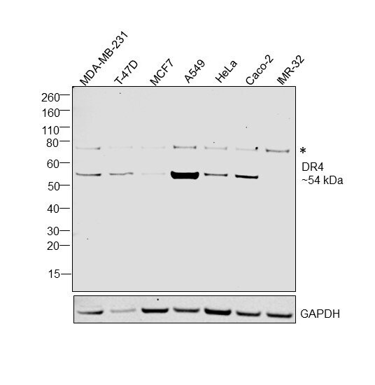
- Western Blot was performed using Anti-DR4 Monoclonal Antibody (32A1380) (Product # MA1-25373) and a 54 kDa band corresponding to Tumor necrosis factor receptor superfamily member 10A was observed across tested cell lines along with an uncharacterised band at ~75 kDa. Membrane enriched extracts (40 µg lysate) of MDA-MB-231 (Lane 1), T-47D (Lane 2), MCF7 (Lane 3), A549 (Lane 4), HeLa (Lane 5), Caco-2 (Lane 6), IMR-32 (Lane 7) were electrophoresed using NuPAGE™ 4-12% Bis-Tris Protein Gel (Product # NP0321BOX). Resolved proteins were then transferred onto a Nitrocellulose membrane (Product # LC2001) by iBlot® 2 Dry Blotting System (Product # IB21001). The Blot was probed with the primary antibody (0.5 µg/mL concentration) and detected by chemiluminescence with Goat anti-Mouse IgG (H+L) Superclonal™ Recombinant Secondary Antibody, HRP (Product # A28177, 1:4000 dilution) using the iBright FL 1000 (Product # A32752). Chemiluminescent detection was performed using Novex® ECL Chemiluminescent Substrate Reagent Kit (Product # WP20005).