Explore
 Validate
 Learn
LearnA02152
antibody from Boster Biological Technology
Targeting: TNFRSF10A
Apo2, CD261, DR4, TRAILR-1, TRAILR1
 Western blot
 Immunocytochemistry
Antibody data
- Antibody Data
- Antigen structure
- References [5]
- Comments [0]
- Validations
- Western blot [1]
Submit
Validation data
Reference
Comment
Report error
- Product number
- A02152 - Provider product page

- Provider
- Boster Biological Technology
- Product name
- Anti-DR4/TNFRSF10A Antibody Picoband™
- Antibody type
- Polyclonal
- Description
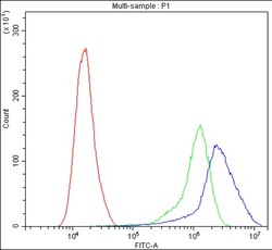
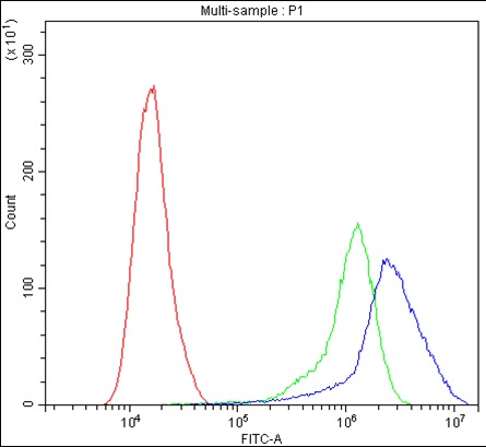
- Polyclonal antibody for DR4/TNFRSF10A detection. Host: Rabbit.Size: 100μg/vial. Tested applications: Flow Cytometry. Reactive species: Human. DR4/TNFRSF10A information: Molecular Weight: 50089 MW; Subcellular Localization: Membrane; Single-pass type I membrane protein; Tissue Specificity: Widely expressed. High levels are found in spleen, peripheral blood leukocytes, small intestine and thymus, but also in K-562 erythroleukemia cells, MCF-7 breast carcinoma cells and activated T-cells.
- Reactivity
- Human, Mouse, Rat
- Host
- Rabbit
- Vial size
- 100μg/vial
- Concentration
- Add 0.2ml of distilled water will yield a concentration of 500ug/ml.
- Storage
- At -20°C for one year. After reconstitution, at 4°C for one month. It can also be aliquoted and stored frozen at -20°C for a longer time. Avoid repeated freezing and thawing.
- Handling
- Add 0.2ml of distilled water will yield a concentration of 500ug/ml.
Submitted references miR-942 decreases TRAIL-induced apoptosis through ISG12a downregulation and is regulated by AKT.
The novel fusion proteins, GnRH-p53 and GnRHIII-p53, expression and their anti-tumor effect.
DJ-1 inhibits TRAIL-induced apoptosis by blocking pro-caspase-8 recruitment to FADD.
Plumbagin enhances TRAIL-mediated apoptosis through up-regulation of death receptor in human melanoma A375 cells.
The occurrence and regional distribution of DR4 on herniated disc cells: a potential apoptosis pathway in lumbar intervertebral disc.
Liu N, Zuo C, Wang X, Chen T, Yang D, Wang J, Zhu H
Oncotarget 2014 Jul 15;5(13):4959-71
Oncotarget 2014 Jul 15;5(13):4959-71
The novel fusion proteins, GnRH-p53 and GnRHIII-p53, expression and their anti-tumor effect.
Jia P, Zhao Y, Wu S, Wu J, Gao S, Tong Y, Wang Y
PloS one 2013;8(11):e79384
PloS one 2013;8(11):e79384
DJ-1 inhibits TRAIL-induced apoptosis by blocking pro-caspase-8 recruitment to FADD.
Fu K, Ren H, Wang Y, Fei E, Wang H, Wang G
Oncogene 2012 Mar 8;31(10):1311-22
Oncogene 2012 Mar 8;31(10):1311-22
Plumbagin enhances TRAIL-mediated apoptosis through up-regulation of death receptor in human melanoma A375 cells.
Li J, Shen Q, Peng R, Chen R, Jiang P, Li Y, Zhang L, Lu J
Journal of Huazhong University of Science and Technology. Medical sciences = Hua zhong ke ji da xue xue bao. Yi xue Ying De wen ban = Huazhong keji daxue xuebao. Yixue Yingdewen ban 2010 Aug;30(4):458-63
Journal of Huazhong University of Science and Technology. Medical sciences = Hua zhong ke ji da xue xue bao. Yi xue Ying De wen ban = Huazhong keji daxue xuebao. Yixue Yingdewen ban 2010 Aug;30(4):458-63
The occurrence and regional distribution of DR4 on herniated disc cells: a potential apoptosis pathway in lumbar intervertebral disc.
Zhang L, Niu T, Yang SY, Lu Z, Chen B
Spine 2008 Feb 15;33(4):422-7
Spine 2008 Feb 15;33(4):422-7
No comments: Submit comment
Supportive validation
- Submitted by
- Boster Biological Technology (provider)
- Main image
- Experimental details
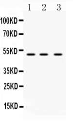
- Western blot analysis of DR4 using anti-DR4 antibody (A02152). Electrophoresis was performed on a 5-20% SDS-PAGE gel at 70V (Stacking gel) / 90V (Resolving gel) for 2-3 hours. The sample well of each lane was loaded with 50ug of sample under reducing conditions. lane 1: rat spleen tissue lysates, lane 2: mouse spleen tissue lysates, lane 3: MCF-7 whole cell lysates. After Electrophoresis, proteins were transferred to a Nitrocellulose membrane at 150mA for 50-90 minutes. Blocked the membrane with 5% Non-fat Milk/ TBS for 1.5 hour at RT. The membrane was incubated with rabbit anti-DR4 antigen affinity purified polyclonal antibody (Catalog # A02152) at 0.5 μg/mL overnight at 4°C, then washed with TBS-0.1%Tween 3 times with 5 minutes each and probed with a goat anti-rabbit IgG-HRP secondary antibody at a dilution of 1:10000 for 1.5 hour at RT. The signal is developed using an Enhanced Chemiluminescent detection (ECL) kit (Catalog # EK1002) with Tanon 5200 system. A specific band was detected for DR4 at approximately 50KD. The expected band size for DR4 is at 50KD.
- Additional image