Explore
 Validate
 Learn
LearnGTX80809
antibody from GeneTex
Targeting: ACTC1
ACTC, CMD1R
 Western blot
 ELISA Immunocytochemistry Immunoprecipitation Immunohistochemistry Chromatin Immunoprecipitation
Antibody data
- Antibody Data
- Antigen structure
- References [1]
- Comments [0]
- Validations
- Western blot [2]
- Immunocytochemistry [2]
- Immunohistochemistry [1]
Submit
Validation data
Reference
Comment
Report error
- Product number
- GTX80809 - Provider product page

- Provider
- GeneTex
- Proper citation
- GeneTex Cat#GTX80809, RRID:AB_626122
- Product name
- Actin antibody [mAbGEa]
- Antibody type
- Monoclonal
- Reactivity
- Human, Mouse, Rat, Bacteria, Bovine, Drosophila, Porcine, Sheep, Xenopus, Yeast, Zebrafish
- Host
- Mouse
Submitted references Curing of the [URE3] prion by Btn2p, a Batten disease-related protein.
Kryndushkin DS, Shewmaker F, Wickner RB
The EMBO journal 2008 Oct 22;27(20):2725-35
The EMBO journal 2008 Oct 22;27(20):2725-35
No comments: Submit comment
Supportive validation
- Submitted by
- GeneTex (provider)
- Main image
- Experimental details
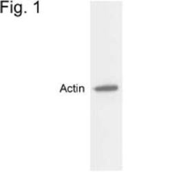
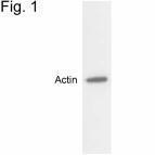
- Western blot detection of actin in MDCK cell extract.

- Submitted by
- GeneTex (provider)
- Main image

- Experimental details
- Western blot of actin in MDCK cell extract using GTX80809.
- Validation comment
- WB
Supportive validation
- Submitted by
- GeneTex (provider)
- Main image

- Experimental details
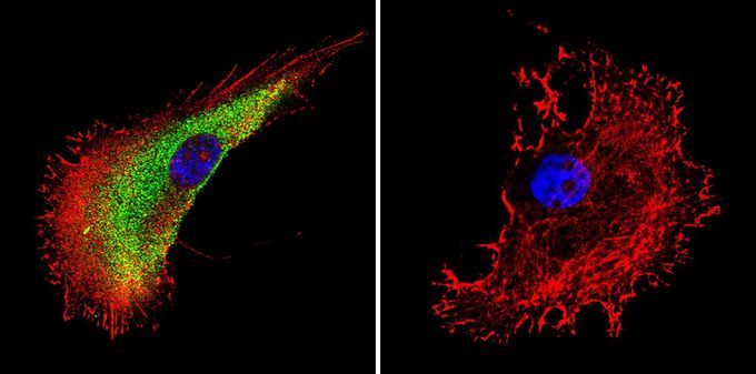
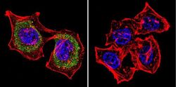
- Immunofluorescent analysis of Actin in A375 cells. Actin staining (green), F-Actin staining with Phalloidin (red) and nuclei with DAPI (blue) is shown. Cells were grown on slides and fixed with formaldehyde prior to staining. Cells were probed without (control) or with Actin antibody [mAbGEa] at a dilution of 1:20 over night at 4 °C, washed with PBS and incubated with a proper secondary antibody. Images were taken at 60X magnification.
- Submitted by
- GeneTex (provider)
- Main image
- Experimental details
- ICC/IF analysis of HeLa cells using Actin antibody at a dilution of 1:20 (green). F-Actin staining with Phalloidin (red) and nuclei with DAPI (blue) is shown. Images were taken at 60X magnification.

Supportive validation
- Submitted by
- GeneTex (provider)
- Main image
- Experimental details
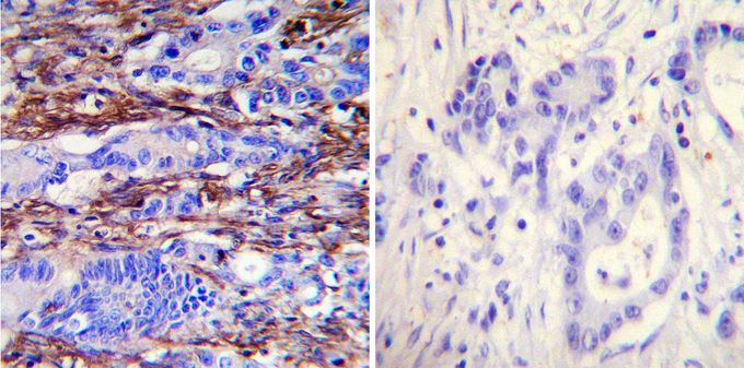
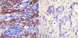
- Immunohistochemistry was performed on cancer biopsies of deparaffinized human colon carcinoma tissues. To expose target proteins, heat induced antigen retrieval was performed using 10mM sodium citrate (pH6.0) buffer, microwaved for 8-15 minutes. Following antigen retrieval tissues were blocked in 3% BSA-PBS for 30 minutes at room temperature. Tissues were then probed at a dilution of 1:1000 with or without Actin antibody [mAbGEa] overnight at 4°C in a humidified chamber. Tissues were washed extensively with PBST and endogenous peroxidase activity was quenched with a peroxidase suppressor. Detection was performed using a biotin-conjμgated secondary antibody and SA-HRP, followed by colorimetric detection using DAB. Tissues were counterstained with hematoxylin and prepped for mounting.