 Explore
 Validate
 Learn
Learn Western blot
Antibody data
- Antibody Data
- Antigen structure
- References [5]
- Comments [0]
- Validations
- Western blot [7]
- Immunocytochemistry [2]
- Immunohistochemistry [3]
- Other assay [3]
Submit
Validation data
Reference
Comment
Report error
- Product number
- PA5-21396 - Provider product page

- Provider
- Invitrogen Antibodies
- Product name
- alpha-Cardiac Actin Polyclonal Antibody
- Antibody type
- Polyclonal
- Antigen
- Recombinant full-length protein
- Description
- Recommended positive controls: Jurkat , Raji, K562, THP-1, NIH-3T3, JC, BCL-1, Rat muscle, PC-12, Rat2. Predicted reactivity: Mouse (100%), Rat (100%), Xenopus laevis (99%), Pig (100%), Chicken (100%), Bovine (100%). Store product as a concentrated solution. Centrifuge briefly prior to opening the vial.
- Reactivity
- Human, Mouse, Rat
- Host
- Rabbit
- Isotype
- IgG
- Vial size
- 100 μL
- Concentration
- 1.39 mg/mL
- Storage
- Store at 4°C short term. For long term storage, store at -20°C, avoiding freeze/thaw cycles.
Submitted references Induction of apoptosis and autosis in cardiomyocytes by the combination of homocysteine and copper via NOX-mediated p62 expression.
Influence of cytoskeleton organization on recombinant protein expression by CHO cells.
Lymphocyte-specific protein 1 regulates mechanosensory oscillation of podosomes and actin isoform-based actomyosin symmetry breaking.
Effects of Ginkgo biloba leaf extract on local renin-angiotensin system through TLR4/NF-κB pathway in cardiac myocyte.
Ascribing novel functions to the sarcomeric protein, myosin binding protein H (MyBPH) in cardiac sarcomere contraction.
Yin R, Wang H, Li C, Wang L, Lai S, Yang X, Hong D, Zhang W
Cell death discovery 2022 Feb 21;8(1):75
Cell death discovery 2022 Feb 21;8(1):75
Influence of cytoskeleton organization on recombinant protein expression by CHO cells.
Pourcel L, Buron F, Arib G, Le Fourn V, Regamey A, Bodenmann I, Girod PA, Mermod N
Biotechnology and bioengineering 2020 Apr;117(4):1117-1126
Biotechnology and bioengineering 2020 Apr;117(4):1117-1126
Lymphocyte-specific protein 1 regulates mechanosensory oscillation of podosomes and actin isoform-based actomyosin symmetry breaking.
Cervero P, Wiesner C, Bouissou A, Poincloux R, Linder S
Nature communications 2018 Feb 6;9(1):515
Nature communications 2018 Feb 6;9(1):515
Effects of Ginkgo biloba leaf extract on local renin-angiotensin system through TLR4/NF-κB pathway in cardiac myocyte.
Jiang H, Qu P
Experimental and therapeutic medicine 2017 Dec;14(6):5857-5862
Experimental and therapeutic medicine 2017 Dec;14(6):5857-5862
Ascribing novel functions to the sarcomeric protein, myosin binding protein H (MyBPH) in cardiac sarcomere contraction.
Mouton J, Loos B, Moolman-Smook JC, Kinnear CJ
Experimental cell research 2015 Feb 15;331(2):338-51
Experimental cell research 2015 Feb 15;331(2):338-51
No comments: Submit comment
Supportive validation
- Submitted by
- Invitrogen Antibodies (provider)
- Main image
- Experimental details
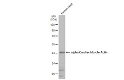
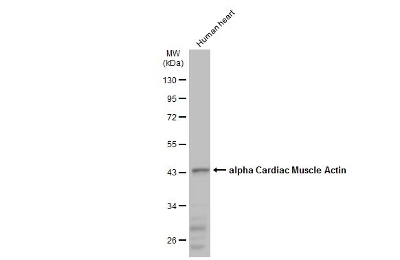
- Western Blot using alpha-Cardiac Actin Polyclonal Antibody (Product # PA5-21396). Human tissue extract (30 µg) was separated by 10% SDS-PAGE, and the membrane was blotted with alpha-Cardiac Actin Polyclonal Antibody (Product # PA5-21396) diluted at 1:1,000. The HRP-conjugated anti-rabbit IgG antibody was used to detect the primary antibody.
- Submitted by
- Invitrogen Antibodies (provider)
- Main image
- Experimental details
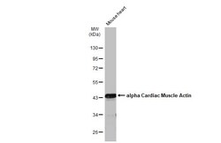
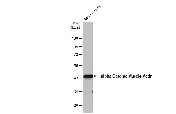
- Western Blot using alpha-Cardiac Actin Polyclonal Antibody (Product # PA5-21396). Mouse tissue extract (50 µg) was separated by 10% SDS-PAGE, and the membrane was blotted with alpha-Cardiac Actin Polyclonal Antibody (Product # PA5-21396) diluted at 1:10,000. The HRP-conjugated anti-rabbit IgG antibody was used to detect the primary antibody.
- Submitted by
- Invitrogen Antibodies (provider)
- Main image
- Experimental details
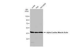
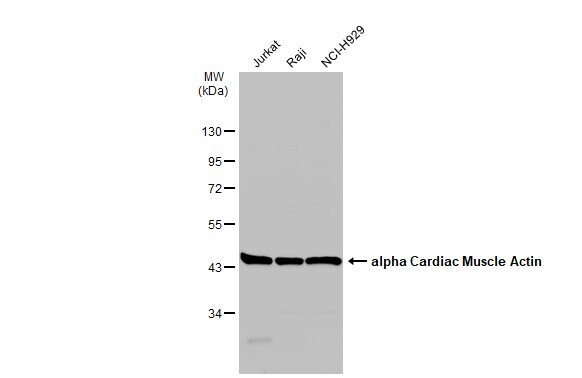
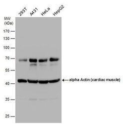
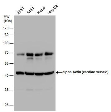
- Western Blot using alpha-Cardiac Actin Polyclonal Antibody (Product # PA5-21396). Various whole cell extracts (30 µg) were separated by 10% SDS-PAGE, and the membrane was blotted with alpha-Cardiac Actin Polyclonal Antibody (Product # PA5-21396) diluted at 1:500. The HRP-conjugated anti-rabbit IgG antibody was used to detect the primary antibody.
- Submitted by
- Invitrogen Antibodies (provider)
- Main image
- Experimental details
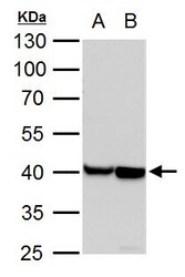
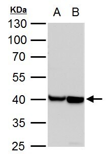
- alpha Actin (cardiac muscle) antibody detects alpha Actin (cardiac muscle) protein by western blot analysis. A. 30 µg PC-12 whole cell lysate/extract. B. 30 µg Rat2 whole cell lysate/extract.10% SDS-PAGE. Alpha Actin (cardiac muscle) antibody alpha-Cardiac Actin Polyclonal Antibody (Product # PA5-21396) dilution: 1:10,000. The HRP-conjugated anti-rabbit IgG antibody was used to detect the primary antibody.
- Submitted by
- Invitrogen Antibodies (provider)
- Main image
- Experimental details
- Western Blot analysis of alpha-Cardiac-Actin was performed by separating 30 µg of various whole cell extracts by 10% SDS-PAGE. Proteins were transferred to a membrane and probed with a alpha-Cardiac Actin Polyclonal Antibody (Product # PA5-21396) at a dilution of 1:500.
- Submitted by
- Invitrogen Antibodies (provider)
- Main image
- Experimental details
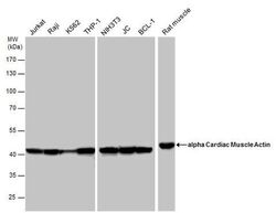
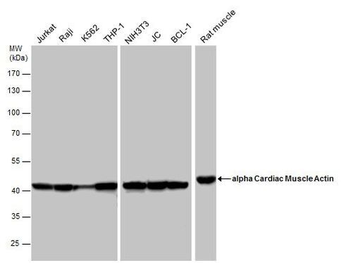
- Western Blot analysis of alpha-Cardiac-Actin was performed by separating 50 µg of various whole cell extracts by 10% SDS-PAGE. Proteins were transferred to a membrane and probed with a alpha-Cardiac Actin Polyclonal Antibody (Product # PA5-21396) at a dilution of 1:10000 and a HRP-conjugated anti-rabbit IgG secondary antibody.
- Submitted by
- Invitrogen Antibodies (provider)
- Main image

- Experimental details
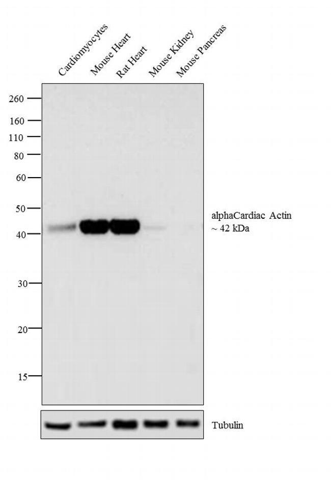
- Western blot analysis was performed on (30 µg lysate) iPSC differentiated to cardiomyocytes (Lane 1), tissue extracts of Mouse Heart (Lane 2), Rat Heart (Lane 3), Mouse Kidney (Lane 4) and Mouse Pancreas (Lane 7). The blot was probed with Anti-alpha-Cardiac Actin Polyclonal Antibody (Product # PA5-21396, 1:5,000 dilution) and detected by chemiluminescence using Goat anti-Rabbit IgG (Heavy Chain) Superclonal™ Secondary Antibody, HRP conjugate (Product # A27036, 0.25 µg/mL, 1:4,000 dilution). A 42 kDa band corresponding to alpha-Cardiac Actin was detected in cardiomyocytes and heart tissues except for Mouse Kidney and Mouse Pancreas which are reported negative for alpha-Cardiac Actin expression.
Supportive validation
- Submitted by
- Invitrogen Antibodies (provider)
- Main image
- Experimental details
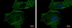
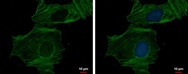
- alpha Actin (cardiac muscle) antibody detects alpha Actin (cardiac muscle) protein at cytoskeleton by immunofluorescent analysis. Sample: HeLa cells were fixed in 0.5% Triton X-100 for 1 min, then ice-cold methanol for 5 min. Green: alpha Actin (cardiac muscle) protein stained by alpha Actin (cardiac muscle) antibody (Product # PA5-21396) diluted at 1:200. Blue: Hoechst 33342 staining.

- Submitted by
- Invitrogen Antibodies (provider)
- Main image

- Experimental details
- alpha Actin (cardiac muscle) antibody detects alpha Actin (cardiac muscle) protein at cytoskeleton by immunofluorescent analysis. Sample: HeLa cells were fixed in 0.5% Triton X-100 for 1 min, then ice-cold methanol for 5 min. Green: alpha Actin (cardiac muscle) protein stained by alpha Actin (cardiac muscle) antibody (Product # PA5-21396) diluted at 1:200. Blue: Hoechst 33342 staining.
Supportive validation
- Submitted by
- Invitrogen Antibodies (provider)
- Main image

- Experimental details
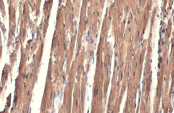
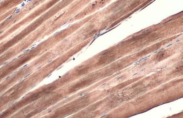
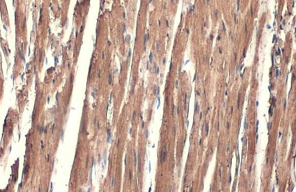
- alpha-Cardiac Actin Polyclonal Antibody detects alpha Cardiac Muscle Actin protein at cytoplasm by immunohistochemical analysis. Sample: Paraffin-embedded mouse heart. alpha Cardiac Muscle Actin stained by alpha-Cardiac Actin Polyclonal Antibody (Product # PA5-21396) diluted at 1:500. Antigen Retrieval: Citrate buffer, pH 6.0, 15 min.
- Submitted by
- Invitrogen Antibodies (provider)
- Main image

- Experimental details
- alpha-Cardiac Actin Polyclonal Antibody detects alpha Cardiac Muscle Actin protein at cytoplasm by immunohistochemical analysis. Sample: Paraffin-embedded mouse muscle. alpha Cardiac Muscle Actin stained by alpha-Cardiac Actin Polyclonal Antibody (Product # PA5-21396) diluted at 1:500. Antigen Retrieval: Citrate buffer, pH 6.0, 15 min.
- Submitted by
- Invitrogen Antibodies (provider)
- Main image

- Experimental details
- alpha-Cardiac Actin Polyclonal Antibody detects alpha Cardiac Muscle Actin protein at cytoplasm by immunohistochemical analysis. Sample: Paraffin-embedded mouse heart. alpha Cardiac Muscle Actin stained by alpha-Cardiac Actin Polyclonal Antibody (Product # PA5-21396) diluted at 1:500. Antigen Retrieval: Citrate buffer, pH 6.0, 15 min.
Supportive validation
- Submitted by
- Invitrogen Antibodies (provider)
- Main image
- Experimental details
- NULL
- Submitted by
- Invitrogen Antibodies (provider)
- Main image
- Experimental details
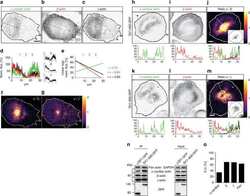
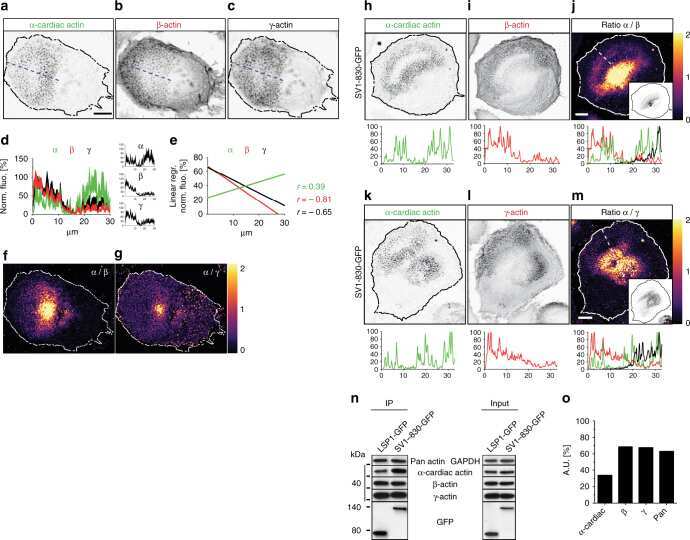
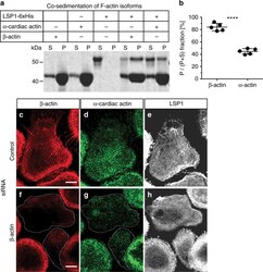
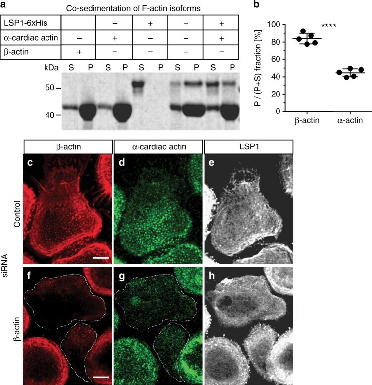
- Fig. 8 Differential subcellular recruitment of LSP1 and supervillin is based on the preferential binding of LSP1 to beta-actin. a Comassie blue staining of gel showing actin cosedimentation assays with pure beta-actin, alpha-cardiac actin, or LSP1 6xHis, alone as control, or in combination with actin isoforms, as indicated. Lanes showing supernatant and pellet fractions are labelled with ""S"" and ""P"", accordingly. Molecular weight is indicated in kDa. b Quantification of copelleted material as ratios of pelleted fraction versus input. Values are given as mean +- S.D.; **** P < 0.0001, two-tailed unpaired t -test. c - h Confocal micrographs of macrophages treated with control siRNA ( c - e ) or beta-actin-specific siRNA ( f - h ), stained for beta-actin ( c , f ), alpha-cardiac actin ( d , g ), and LSP1 ( e , h ). (Note: staining conditions required especially for alpha-cardiac actin are not optimal for staining of LSP1.) Scale bars: 10 um. For specific values, see Supplementary Data 1
- Submitted by
- Invitrogen Antibodies (provider)
- Main image
- Experimental details
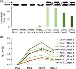
- 3 Characterization of the productivity of ACTC1-overexpressing cells. A trastuzumab-expressing line was stably retransfected with the CHO ACTC1 or with an empty expression vector, and cell clones were isolated for further analysis. (a) Immunoblots of total protein extracts labeled with ACTC1 or GAPDH mouse antibodies. The histogram shows the ratio of the ACTC1 signal relative to that of GAPDH, as assessed using the ImageJ. Ponceau red-stained membranes are shown in Figure S3a. (b) Secreted IgG titers in culture supernatants were assessed by double sandwich ELISA over 13 days of fed-batch cultures. CHO, Chinese hamster ovary; ELISA, enzyme-linked immunosorbent assay; IgG, immunoglobulin gamma [Color figure can be viewed at wileyonlinelibrary.com]
