Explore
 Validate
 Learn
Learn Western blot
Antibody data
- Antibody Data
- Antigen structure
- References [1]
- Comments [0]
- Validations
- Western blot [3]
- Immunocytochemistry [2]
- Immunohistochemistry [1]
- Other assay [1]
Submit
Validation data
Reference
Comment
Report error
- Product number
- PA5-11570 - Provider product page

- Provider
- Invitrogen Antibodies
- Product name
- Actin Polyclonal Antibody
- Antibody type
- Polyclonal
- Antigen
- Synthetic peptide
- Description
- This antibody is predicted to react with bovine, chicken, canine, equine, hamster, porcine, non-human primate, rat, rabbit and Xenopus, yeast and zebrafish based on sequence homology.
- Reactivity
- Human, Mouse
- Host
- Rabbit
- Vial size
- 400 µL
- Concentration
- 1.5 mg/mL
- Storage
- -20° C, Avoid Freeze/Thaw Cycles
Submitted references Bisphenol A, Bisphenol F, and Bisphenol S: The Bad and the Ugly. Where Is the Good?
Fouyet S, Olivier E, Leproux P, Dutot M, Rat P
Life (Basel, Switzerland) 2021 Apr 3;11(4)
Life (Basel, Switzerland) 2021 Apr 3;11(4)
No comments: Submit comment
Supportive validation
- Submitted by
- Invitrogen Antibodies (provider)
- Main image
- Experimental details
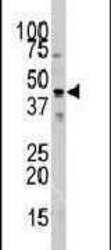
- Western blot analysis using an Actin polyclonal antibody (Product # PA5-11570) in mouse testis tissue lysates (35 µg per lane).
- Submitted by
- Invitrogen Antibodies (provider)
- Main image

- Experimental details
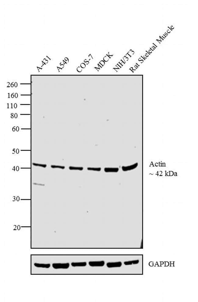
- Western blot analysis was performed on whole cell extracts (30 µg lysate) of A-431 (Lane 1), A549 (Lane 2), COS-7 (Lane 3), MDCK (Lane 4), NIH/3T3 (Lane 5) tissue extracts of and Rat Skeletal Muscle (Lane 6). The blot was probed with Actin Polyclonal Antibody (Product # PA5-11570, 1:1000 dilution) and detected by chemiluminescence using Goat anti-Rabbit IgG (H+L) Superclonal™ Secondary Antibody, HRP conjugate (Product # A27036, 0.25 µg/mL, 1:4000 dilution). A 42 kDa band corresponding to Actin was observed across the cell lines and tissues tested.
- Submitted by
- Invitrogen Antibodies (provider)
- Main image

- Experimental details
- Western blot analysis was performed on whole cell extracts (30 µg lysate) of A-431 (Lane 1), A549 (Lane 2), COS-7 (Lane 3), MDCK (Lane 4), NIH/3T3 (Lane 5) tissue extracts of and Rat Skeletal Muscle (Lane 6). The blot was probed with Actin Polyclonal Antibody (Product # PA5-11570, 1:1000 dilution) and detected by chemiluminescence using Goat anti-Rabbit IgG (H+L) Superclonal™ Secondary Antibody, HRP conjugate (Product # A27036, 0.25 µg/mL, 1:4000 dilution). A 42 kDa band corresponding to Actin was observed across the cell lines and tissues tested.

Supportive validation
- Submitted by
- Invitrogen Antibodies (provider)
- Main image

- Experimental details
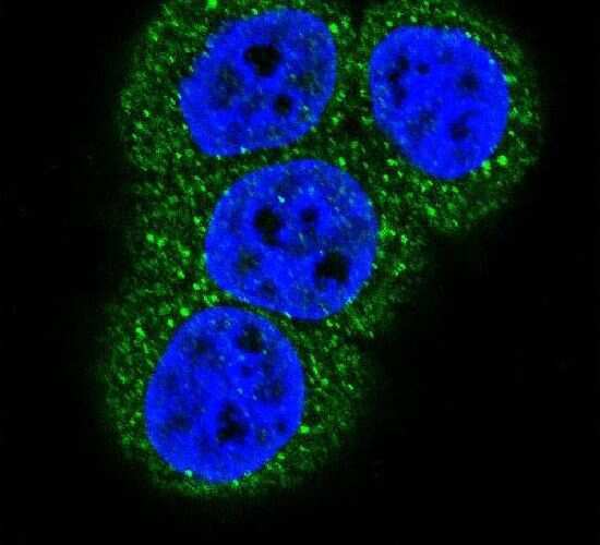
- Immunofluorescent analysis of HeLa cells using an Actin polyclonal antibody (Product # PA5-11570) at a dilution of 1:10-50, followed by a fluor-conjugated goat anti-rabbit secondary antibody (green). Nuclei were stained with DAPI (blue).
- Submitted by
- Invitrogen Antibodies (provider)
- Main image

- Experimental details
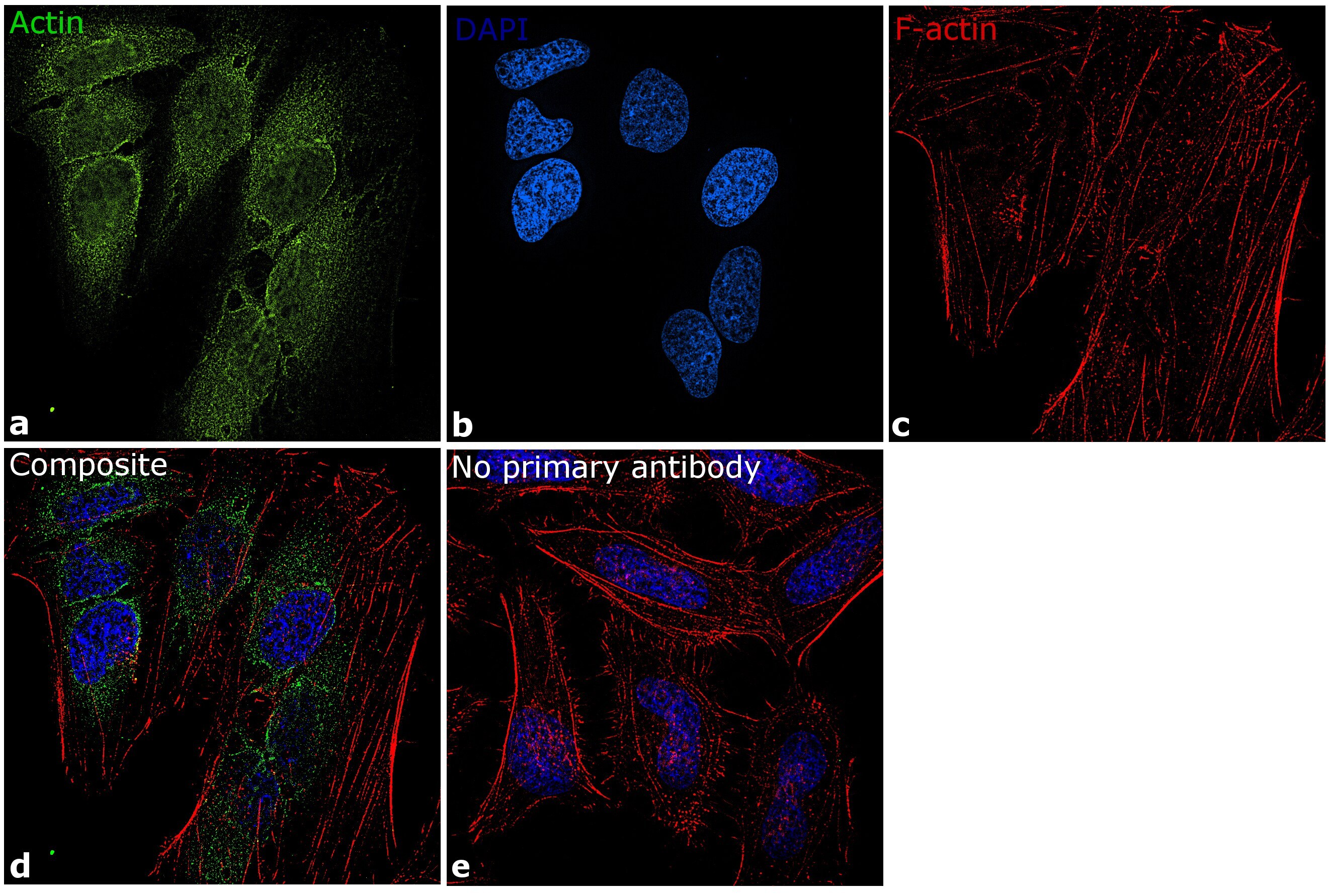
- Immunofluorescence analysis of Actin was performed using 70% confluent log phase HeLa cells. The cells were fixed with 4% paraformaldehyde for 10 minutes, permeabilized with 0.1% Triton™ X-100 for 10 minutes, and blocked with 1% BSA for 1 hour at room temperature. The cells were labeled with Actin Polyclonal Antibody (Product # PA5-11570) at 1:50 dilution in 0.1% BSA and incubated overnight at 4 degree and then labeled with Goat anti-Rabbit IgG (H+L) Superclonal™ Secondary Antibody, Alexa Fluor® 488 conjugate (Product # A27034) at a dilution of 1:2000 for 45 minutes at room temperature (Panel a: green). Nuclei (Panel b: blue) were stained with SlowFade® Gold Antifade Mountant with DAPI (Product # S36938). F-actin (Panel c: red) was stained with Rhodamine Phalloidin (Product # R415, 1:300). Panel d represents the merged image showing cytoskeletal localization. Panel e represents control cells with no primary antibody to assess background. The images were captured at 60X magnification.
Supportive validation
- Submitted by
- Invitrogen Antibodies (provider)
- Main image
- Experimental details
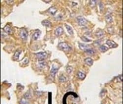
- Immunohistochemical analysis of formalin-fixed, paraffin-embedded human lung carcinoma tissue using an Actin polyclonal antibody (Product # PA5-11570), followed by HRP-conjugated secondary antibody and DAB staining.

Supportive validation
- Submitted by
- Invitrogen Antibodies (provider)
- Main image

- Experimental details
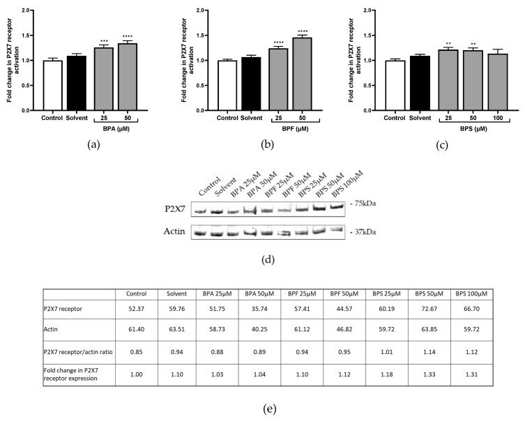
- Figure 3 ( a - c ) P2X7 receptor activation was evaluated after ( a ) BPA, ( b ) BPF, and ( c ) BPS incubation for 72 h in JEG-Tox cells. Data correspond to the mean +-SEM of four independent experiments. The significance thresholds were **** p < 0.0001, *** p < 0.001, and ** p < 0.01 compared to the control. ( d , e ) P2X7 receptor expression was assessed after BPA, BPF, and BPS incubation for 72 h in JEG-Tox cells ( d ) using the Western blot technique, and ( e ) quantitative analysis was performed using ImageJ software. beta-actin was used as a control.