 Explore
 Validate
 Learn
Learn Western blot
 Immunoprecipitation
Antibody data
- Antibody Data
- Antigen structure
- References [6]
- Comments [0]
- Validations
- Western blot [2]
- Immunohistochemistry [1]
Submit
Validation data
Reference
Comment
Report error
- Product number
- NB100-1828 - Provider product page

- Provider
- Novus Biologicals
- Proper citation
- Novus Cat#NB100-1828, RRID:AB_10001847
- Product name
- Mouse Monoclonal NKX3.1 Antibody
- Antibody type
- Monoclonal
- Description
- Protein G purified.
- Reactivity
- Human, Mouse, Rat
- Host
- Mouse
- Isotype
- IgG
- Vial size
- 0.1 ml
- Concentration
- 2.3 mg/ml
- Storage
- Store at 4C short term. Aliquot and store at -20C long term. Avoid freeze-thaw cycles.
Submitted references Epigenetic silencing of SALL2 confers tamoxifen resistance in breast cancer.
TMPRSS2-ERG Controls Luminal Epithelial Lineage and Antiandrogen Sensitivity in PTEN and TP53-Mutated Prostate Cancer.
p300 acetyltransferase regulates androgen receptor degradation and PTEN-deficient prostate tumorigenesis.
Nkx3.1 functions as para-transcription factor to regulate gene expression and cell proliferation in non-cell autonomous manner.
A critical role for p27kip1 gene dosage in a mouse model of prostate carcinogenesis.
Cooperativity of Nkx3.1 and Pten loss of function in a mouse model of prostate carcinogenesis.
Ye L, Lin C, Wang X, Li Q, Li Y, Wang M, Zhao Z, Wu X, Shi D, Xiao Y, Ren L, Jian Y, Yang M, Ou R, Deng G, Ouyang Y, Chen X, Li J, Song L
EMBO molecular medicine 2019 Dec;11(12):e10638
EMBO molecular medicine 2019 Dec;11(12):e10638
TMPRSS2-ERG Controls Luminal Epithelial Lineage and Antiandrogen Sensitivity in PTEN and TP53-Mutated Prostate Cancer.
Blee AM, He Y, Yang Y, Ye Z, Yan Y, Pan Y, Ma T, Dugdale J, Kuehn E, Kohli M, Jimenez R, Chen Y, Xu W, Wang L, Huang H
Clinical cancer research : an official journal of the American Association for Cancer Research 2018 Sep 15;24(18):4551-4565
Clinical cancer research : an official journal of the American Association for Cancer Research 2018 Sep 15;24(18):4551-4565
p300 acetyltransferase regulates androgen receptor degradation and PTEN-deficient prostate tumorigenesis.
Zhong J, Ding L, Bohrer LR, Pan Y, Liu P, Zhang J, Sebo TJ, Karnes RJ, Tindall DJ, van Deursen J, Huang H
Cancer research 2014 Mar 15;74(6):1870-1880
Cancer research 2014 Mar 15;74(6):1870-1880
Nkx3.1 functions as para-transcription factor to regulate gene expression and cell proliferation in non-cell autonomous manner.
Zhou J, Qin L, Tien JC, Gao L, Chen X, Wang F, Hsieh JT, Xu J
The Journal of biological chemistry 2012 May 18;287(21):17248-56
The Journal of biological chemistry 2012 May 18;287(21):17248-56
A critical role for p27kip1 gene dosage in a mouse model of prostate carcinogenesis.
Gao H, Ouyang X, Banach-Petrosky W, Borowsky AD, Lin Y, Kim M, Lee H, Shih WJ, Cardiff RD, Shen MM, Abate-Shen C
Proceedings of the National Academy of Sciences of the United States of America 2004 Dec 7;101(49):17204-9
Proceedings of the National Academy of Sciences of the United States of America 2004 Dec 7;101(49):17204-9
Cooperativity of Nkx3.1 and Pten loss of function in a mouse model of prostate carcinogenesis.
Kim MJ, Cardiff RD, Desai N, Banach-Petrosky WA, Parsons R, Shen MM, Abate-Shen C
Proceedings of the National Academy of Sciences of the United States of America 2002 Mar 5;99(5):2884-9
Proceedings of the National Academy of Sciences of the United States of America 2002 Mar 5;99(5):2884-9
No comments: Submit comment
Supportive validation
- Submitted by
- Novus Biologicals (provider)
- Main image
- Experimental details
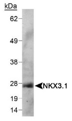
- Western Blot: Nkx3.1 Antibody (0361) [NB100-1828] - Detection of NKX3.1 in mouse testis lysate using NB 100-1828. ECL detection 1 minute.

- Submitted by
- Novus Biologicals (provider)
- Main image

- Experimental details
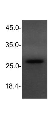
- Western Blot: NKX3.1 Antibody (0361) [NB100-1828] - analysis of NKX3.1 in HeLa cell lysate using anti-NKX3.1 antibody. Image from verified customer review.
Supportive validation
- Submitted by
- Novus Biologicals (provider)
- Main image
- Experimental details
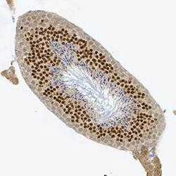
- Immunohistochemistry-Paraffin: NKX3.1 Antibody (0361) [NB100-1828] - NKX3.1 was detected in immersion fixed paraffin-embedded sections of mouse testis using Mouse Anti-Mouse NKX3.1 (0361) Monoclonal Antibody (Catalog # NB100-1828) at 1:300 for 1 hour at room temperature followed by incubation with the Anti-Mouse IgG VisUCyte™ HRP Polymer Antibody (Catalog # VC001). Tissue was stained using DAB (brown) and counterstained with hematoxylin (blue). Specific staining was localized to the nuclei in sperm cells.
