 Explore
 Validate
 Learn
Learn Western blot
Antibody data
- Antibody Data
- Antigen structure
- References [0]
- Comments [0]
- Validations
- Western blot [2]
- Immunocytochemistry [2]
- Flow cytometry [1]
Submit
Validation data
Reference
Comment
Report error
- Product number
- TA504135 - Provider product page

- Provider
- OriGene
- Proper citation
- OriGene Cat#TA504135, RRID:AB_2622418
- Product name
- CST4 (Cystatin S) mouse monoclonal antibody, clone OTI2H10 (formerly 2H10)
- Antibody type
- Monoclonal
- Description
- CST4 (Cystatin S) mouse monoclonal antibody, clone OTI2H10 (formerly 2H10)
- Host
- Mouse
- Conjugate
- Unconjugated
- Epitope
- CST4
- Isotype
- IgG
- Antibody clone number
- OTI2H10
- Vial size
- 100 µl
- Concentration
- NULL
No comments: Submit comment
Supportive validation
- Submitted by
- OriGene (provider)
- Main image
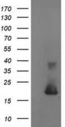
- Experimental details
- HEK293T cells were transfected with the pCMV6-ENTRY control (Left lane) or pCMV6-ENTRY CST4 (RC209349, Right lane) cDNA for 48 hrs and lysed. Equivalent amounts of cell lysates (5 ug per lane) were separated by SDS-PAGE and immunoblotted with anti-CST4.
- Validation comment
- WB
- Submitted by
- OriGene (provider)
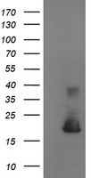
- Main image
- Experimental details
- Western blot analysis of extracts (10ug) from 1 cell line by using anti-CST4 monoclonal antibody at 1:200 dilution.
- Validation comment
- WB

Supportive validation
- Submitted by
- OriGene (provider)
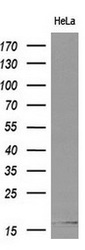
- Main image
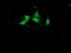
- Experimental details
- Immunofluorescent staining of HeLa cells using anti-CST4 mouse monoclonal antibody (TA504135).
- Validation comment
- IF

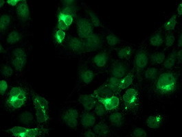
- Submitted by
- OriGene (provider)
- Main image

- Experimental details
- Anti-CST4 mouse monoclonal antibody (TA504135) immunofluorescent staining of COS7 cells transiently transfected by pCMV6-ENTRY CST4(RC209349).
- Validation comment
- IF
Supportive validation
- Submitted by
- OriGene (provider)
- Main image
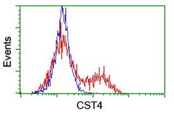
- Experimental details
- HEK293T cells transfected with either RC209349 overexpress plasmid(Red) or empty vector control plasmid(Blue) were immunostained by anti-CST4 antibody(TA504135), and then analyzed by flow cytometry.
- Validation comment
- FC
