 Explore
 Validate
 Learn
Learn Western blot
Antibody data
- Antibody Data
- Antigen structure
- References [2]
- Comments [0]
- Validations
- Western blot [1]
- Immunocytochemistry [2]
- Immunohistochemistry [2]
- Flow cytometry [2]
Submit
Validation data
Reference
Comment
Report error
- Product number
- TA503075 - Provider product page

- Provider
- OriGene
- Proper citation
- OriGene Cat#TA503075, RRID:AB_11139727
- Product name
- RGS5 mouse monoclonal antibody, clone OTI1C1 (formerly 1C1)
- Antibody type
- Monoclonal
- Description
- RGS5 mouse monoclonal antibody, clone OTI1C1 (formerly 1C1)
- Host
- Mouse
- Conjugate
- Unconjugated
- Epitope
- RGS5
- Isotype
- IgG
- Antibody clone number
- OTI1C1
- Vial size
- 100 µl
- Concentration
- 1 mg/ml
Submitted references Brain pericytes acquire a microglial phenotype after stroke.
Lipid rafts are required for signal transduction by angiotensin II receptor type 1 in neonatal glomerular mesangial cells.
Özen I, Deierborg T, Miharada K, Padel T, Englund E, Genové G, Paul G
Acta neuropathologica 2014 Sep;128(3):381-96
Acta neuropathologica 2014 Sep;128(3):381-96
Lipid rafts are required for signal transduction by angiotensin II receptor type 1 in neonatal glomerular mesangial cells.
Adebiyi A, Soni H, John TA, Yang F
Experimental cell research 2014 May 15;324(1):92-104
Experimental cell research 2014 May 15;324(1):92-104
No comments: Submit comment
Supportive validation
- Submitted by
- OriGene (provider)
- Main image
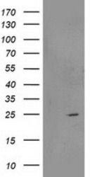
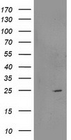
- Experimental details
- HEK293T cells were transfected with the pCMV6-ENTRY control (Left lane) or pCMV6-ENTRY RGS5 (RC206857, Right lane) cDNA for 48 hrs and lysed. Equivalent amounts of cell lysates (5 ug per lane) were separated by SDS-PAGE and immunoblotted with anti-RGS5.
- Validation comment
- WB
Supportive validation
- Submitted by
- OriGene (provider)
- Main image
- Experimental details
- Figure from citation: Immunofluorescence of RGS5 protein level by using anti-RGS5 antibody in primary neonatal pig GMCs.
- Validation comment
- IF
- Submitted by
- OriGene (provider)
- Main image
- Experimental details
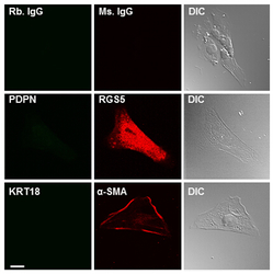
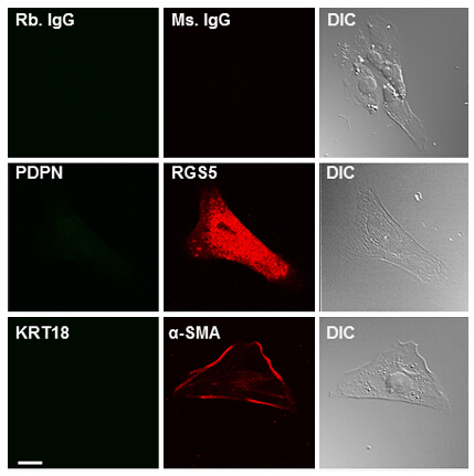
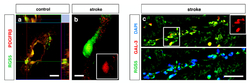
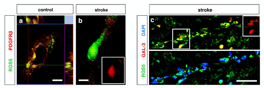
- Figure from citation: Immunofluorescence of RGS5 protein level by using anti-RGS5 antibody in human brain pericytes. Dilution: 1:200
- Validation comment
- IF
Supportive validation
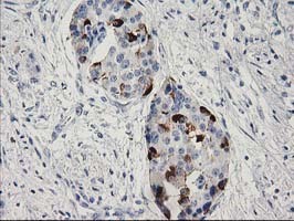
- Submitted by
- OriGene (provider)
- Main image
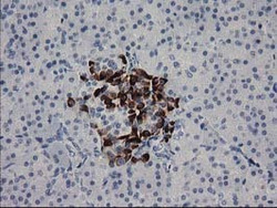
- Experimental details
- Immunohistochemical staining of paraffin-embedded Human pancreas tissue within the normal limits using anti-RGS5 mouse monoclonal antibody. (Heat-induced epitope retrieval by 10mM citric buffer, pH6.0, 100C for 10min, TA503075)
- Validation comment
- IHC
- Submitted by
- OriGene (provider)
- Main image
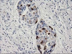
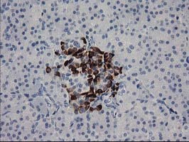
- Experimental details
- Immunohistochemical staining of paraffin-embedded Carcinoma of Human pancreas tissue using anti-RGS5 mouse monoclonal antibody. (Heat-induced epitope retrieval by 10mM citric buffer, pH6.0, 100C for 10min, TA503075)
- Validation comment
- IHC
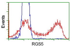
Supportive validation
- Submitted by
- OriGene (provider)
- Main image
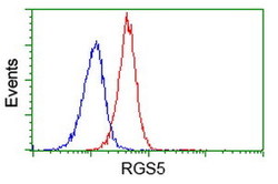
- Experimental details
- Flow cytometric Analysis of Jurkat cells, using anti-RGS5 antibody(TA503075),(Red), compared to a nonspecific negative control antibody,(Blue).
- Validation comment
- FC
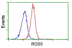
- Submitted by
- OriGene (provider)
- Main image
- Experimental details
- HEK293T cells transfected with either RC206857 overexpress plasmid(Red) or empty vector control plasmid(Blue) were immunostained by anti-RGS5 antibody(TA503075), and then analyzed by flow cytometry.
- Validation comment
- FC
