 Explore
 Validate
 Learn
Learn Western blot
Antibody data
- Antibody Data
- Antigen structure
- References [2]
- Comments [0]
- Validations
- Western blot [2]
- Immunohistochemistry [1]
Submit
Validation data
Reference
Comment
Report error
- Product number
- AF5890 - Provider product page

- Provider
- R&D Systems
- Product name
- Mouse Contactin-6 Antibody
- Antibody type
- Polyclonal
- Description
- Immunogen affinity purified. Detects mouse Contactin-6 in direct ELISAs and Western blots. In direct ELISAs, approximately 50% cross-reactivity with recombinant human (rh) Contactin-6 is observed, and less than 5% cross-reactivity with recombinant mouse (rm) Contactin-3, rmContactin-4, rmContactin-5, and rhTAG-1 is observed.
- Reactivity
- Mouse
- Host
- Sheep
- Conjugate
- Unconjugated
- Antigen sequence
Q9JMB8- Isotype
- IgG
- Vial size
- 100 ug
- Concentration
- LYOPH
- Storage
- Use a manual defrost freezer and avoid repeated freeze-thaw cycles. 12 months from date of receipt, -20 to -70 °C as supplied. 1 month, 2 to 8 °C under sterile conditions after reconstitution. 6 months, -20 to -70 °C under sterile conditions after reconstitution.
Submitted references Association of Cell Adhesion Molecules Contactin-6 and Latrophilin-1 Regulates Neuronal Apoptosis.
Loss of NB-3 aggravates cerebral ischemia by impairing neuron survival and neurite growth.
Zuko A, Oguro-Ando A, Post H, Taggenbrock RL, van Dijk RE, Altelaar AF, Heck AJ, Petrenko AG, van der Zwaag B, Shimoda Y, Pasterkamp RJ, Burbach JP
Frontiers in molecular neuroscience 2016;9:143
Frontiers in molecular neuroscience 2016;9:143
Loss of NB-3 aggravates cerebral ischemia by impairing neuron survival and neurite growth.
Huang X, Sun J, Zhao T, Wu KW, Watanabe K, Xiao ZC, Zhu LL, Fan M
Stroke 2011 Oct;42(10):2910-6
Stroke 2011 Oct;42(10):2910-6
No comments: Submit comment
Supportive validation
- Submitted by
- R&D Systems (provider)
- Main image
- Experimental details
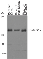
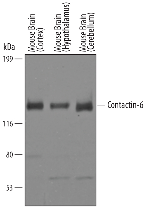
- Detection of Mouse Contactin-6 by Western Blot. Western blot shows lysates of mouse brain (cortex) tissue, mouse brain (hypothalamus) tissue, and mouse brain (cerebellum) tissue. PVDF Membrane was probed with 1 µg/mL of Sheep Anti-Mouse Contactin-6 Antigen Affinity-purified Polyclonal Antibody (Catalog # AF5890) followed by HRP-conjugated Anti-Sheep IgG Secondary Antibody (Catalog # HAF016). A specific band was detected for Contactin-6 at approximately 130 kDa (as indicated). This experiment was conducted under reducing conditions and using Immunoblot Buffer Group 8.
- Submitted by
- R&D Systems (provider)
- Main image
- Experimental details
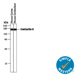
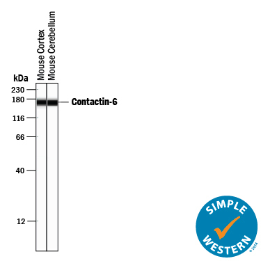
- Detection of Mouse Contactin-6 by Simple WesternTM. Simple Western lane view shows lysates of mouse brain (cortex) tissue and mouse brain (cerebellum) tissue, loaded at 0.2 mg/mL. A specific band was detected for Contactin-6 at approximately 168 kDa (as indicated) using 10 µg/mL of Sheep Anti-Mouse Contactin-6 Antigen Affinity-purified Polyclonal Antibody (Catalog # AF5890) followed by 1:50 dilution of HRP-conjugated Anti-Sheep IgG Secondary Antibody (Catalog # HAF016). This experiment was conducted under reducing conditions and using the 12-230 kDa separation system.
Supportive validation
- Submitted by
- R&D Systems (provider)
- Main image
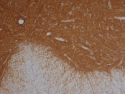
- Experimental details
- Contactin-6 in Mouse Spinal Cord. Contactin-6 was detected in perfusion fixed frozen sections of adult mouse spinal cord using Sheep Anti-Mouse Contactin-6 Antigen Affinity-purified Poly-clonal Antibody (Catalog # AF5890) at 15 µg/mL overnight at 4 °C. Tissue was stained using the Anti-Sheep HRP-DAB Cell & Tissue Staining Kit (brown; Catalog # CTS019) and counter-stained with hematoxylin (blue). Specific staining was localized to gray matter. View our protocol for Chromogenic IHC Staining of Frozen Tissue Sections.
