 Explore
 Validate
 Learn
Learn Western blot
 Immunocytochemistry
Antibody data
- Antibody Data
- Antigen structure
- References [5]
- Comments [0]
- Validations
- Western blot [2]
- Immunohistochemistry [1]
Submit
Validation data
Reference
Comment
Report error
- Product number
- NB100-56368 - Provider product page

- Provider
- Novus Biologicals
- Proper citation
- Novus Cat#NB100-56368, RRID:AB_838643
- Product name
- Rabbit Polyclonal Noxa Antibody
- Antibody type
- Polyclonal
- Description
- Protein G purified.
- Reactivity
- Human, Mouse
- Host
- Rabbit
- Isotype
- IgG
- Vial size
- 0.1 mg
- Concentration
- 1 mg/ml
- Storage
- Store at 4C short term. Aliquot and store at -20C long term. Avoid freeze-thaw cycles.
Submitted references Noxa couples lysosomal membrane permeabilization and apoptosis during oxidative stress.
Molecular regulation of DNA damage-induced apoptosis in neurons of cerebral cortex.
Paraquat neurotoxicity is mediated by a Bak-dependent mechanism.
Apoptosis mediated by p53 in rat neural AF5 cells following treatment with hydrogen peroxide and staurosporine.
Bim and Noxa are candidates to mediate the deleterious effect of the NF-kappa B subunit RelA in cerebral ischemia.
Eno CO, Zhao G, Venkatanarayan A, Wang B, Flores ER, Li C
Free radical biology & medicine 2013 Dec;65:26-37
Free radical biology & medicine 2013 Dec;65:26-37
Molecular regulation of DNA damage-induced apoptosis in neurons of cerebral cortex.
Martin LJ, Liu Z, Pipino J, Chestnut B, Landek MA
Cerebral cortex (New York, N.Y. : 1991) 2009 Jun;19(6):1273-93
Cerebral cortex (New York, N.Y. : 1991) 2009 Jun;19(6):1273-93
Paraquat neurotoxicity is mediated by a Bak-dependent mechanism.
Fei Q, McCormack AL, Di Monte DA, Ethell DW
The Journal of biological chemistry 2008 Feb 8;283(6):3357-64
The Journal of biological chemistry 2008 Feb 8;283(6):3357-64
Apoptosis mediated by p53 in rat neural AF5 cells following treatment with hydrogen peroxide and staurosporine.
McNeill-Blue C, Wetmore BA, Sanchez JF, Freed WJ, Merrick BA
Brain research 2006 Sep 27;1112(1):1-15
Brain research 2006 Sep 27;1112(1):1-15
Bim and Noxa are candidates to mediate the deleterious effect of the NF-kappa B subunit RelA in cerebral ischemia.
Inta I, Paxian S, Maegele I, Zhang W, Pizzi M, Spano P, Sarnico I, Muhammad S, Herrmann O, Inta D, Baumann B, Liou HC, Schmid RM, Schwaninger M
The Journal of neuroscience : the official journal of the Society for Neuroscience 2006 Dec 13;26(50):12896-903
The Journal of neuroscience : the official journal of the Society for Neuroscience 2006 Dec 13;26(50):12896-903
No comments: Submit comment
Supportive validation
- Submitted by
- Novus Biologicals (provider)
- Main image
- Experimental details
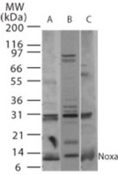
- Western Blot: Noxa Antibody [NB100-56368] - Western blot analysis of Noxa in A) human, B) mouse, and C) rat thymus tissue using Noxa antibody NB100-56368 at 2 ug/mL.

- Submitted by
- Novus Biologicals (provider)
- Main image
- Experimental details
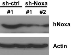
- Western Blot: Noxa Antibody [NB100-56368] - Noxa expression was stably reduced in HCT116 cells by shRNA. Western blot image submitted by a verified customer review.

Supportive validation
- Submitted by
- Novus Biologicals (provider)
- Main image
- Experimental details
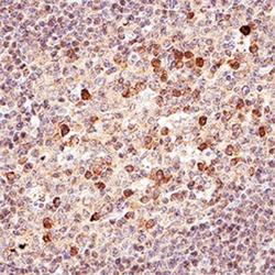
- Immunohistochemistry-Paraffin: Noxa Antibody [NB100-56368] - IHC analysis of formalin fixed paraffin-embedded (FFPE) human tonsil using Noxa antibody at 1:50 on a Bond Rx autostainer (Leica Biosystems). The assay involved 20 minutes of heat induced antigen retrieval (HIER) using 10 mM sodium citrate buffer (pH 6.0) and endogenous peroxidase quenching with peroxide block. The sections were incubated with primary antibody for 30 minutes and Bond Polymer Refine Detection (Leica Biosystems) with DAB was used for signal development followed by counterstaining with hematoxylin. Whole slide scanning and capturing of representative images was performed using Aperio AT2 (Leica Biosystems). Staining was performed by Histowiz.
