 Explore
 Validate
 Learn
Learn Western blot
Antibody data
- Antibody Data
- Antigen structure
- References [0]
- Comments [0]
- Validations
- Western blot [5]
- Immunocytochemistry [1]
- Flow cytometry [1]
Submit
Validation data
Reference
Comment
Report error
- Product number
- 701158 - Provider product page

- Provider
- Invitrogen Antibodies
- Product name
- FABP4 Recombinant Rabbit Monoclonal Antibody (2H3L2)
- Antibody type
- Monoclonal
- Antigen
- Recombinant full-length protein
- Reactivity
- Human, Mouse, Rat
- Host
- Rabbit
- Isotype
- IgG
- Antibody clone number
- 2H3L2
- Vial size
- 100 µg
- Concentration
- 0.5 mg/mL
- Storage
- Store at 4°C short term. For long term storage, store at -20°C, avoiding freeze/thaw cycles.
No comments: Submit comment
Supportive validation
- Submitted by
- Invitrogen Antibodies (provider)
- Main image
- Experimental details
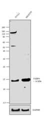
- Western blot analysis was performed on membrane enriched extracts (30 µg lysate) of 3T3-L1 (Lane 1) and SH-SY5Y (Lane 2). The blots were probed with Anti-FABP4 Rabbit Monoclonal Antibody (Product # 701158, 2 µg/mL) and detected by chemiluminescence using Goat anti-Rabbit IgG (H+L) Superclonal™ Secondary Antibody, HRP conjµgate (Product # A27036, 0.4 µg/mL, 1:2,500 dilution). A 14 kDa band corresponding to FABP4 was observed across the cell lines tested. Known quantity of protein samples were electrophoresed using Novex® NuPAGE® 4-12 % Bis-Tris gel (Product # NP0321BOX), XCell SureLock™ Electrophoresis System (Product # EI0002) and Novex® Sharp Pre-Stained Protein Standard (Product # LC5800). Resolved proteins were then transferred onto a nitrocellulose membrane with iBlot® 2 Dry Blotting System (Product # IB21001). The membrane was probed with the relevant primary and secondary Antibody following blocking with 5 % skimmed milk. Chemiluminescent detection was performed using Pierce™ ECL Western Blotting Substrate (Product # 32106).

- Submitted by
- Invitrogen Antibodies (provider)
- Main image
- Experimental details
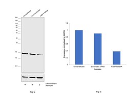
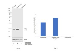
- Knockdown of Fatty acid-binding protein, adipocyte was achieved by transfecting differentiated 3T3-L1 cells to adipocytes with Fatty acid-binding protein, adipocyte specific siRNAs (Silencer® select Product # s62398, s62400). Western blot analysis (Fig. a) was performed using Whole cell extracts from the Fatty acid-binding protein, adipocyte knockdown cells (lane 3), non-targeting scrambled siRNA transfected cells (lane 2) and untransfected cells (lane 1). The blot was probed with FABP4 Recombinant Rabbit Monoclonal Antibody (2H3L2) (Product # 701158, 2 µg/mL ) and Goat anti-Rabbit IgG (H+L) Superclonal™ Recombinant Secondary Antibody, HRP (Product # A27036, 1:20,000 dilution). Densitometric analysis of this western blot is shown in histogram (Fig. b). Decrease in signal upon siRNA mediated knock down confirms that antibody is specific to Fatty acid-binding protein, adipocyte.

- Submitted by
- Invitrogen Antibodies (provider)
- Main image
- Experimental details
- Knockdown of Fatty acid-binding protein, adipocyte was achieved by transfecting 3T3-L1 cells with Fatty acid-binding protein specific siRNAs (Silencer® select Product # s62398, s62400) and then the cells were differentiated to adipocytes. Western blot analysis (Fig. a) was performed using Whole cell extracts from the Fatty acid-binding protein, adipocyte knockdown cells (lane 3), non-targeting scrambled siRNA transfected cells (lane 2) and untransfected cells (lane 1). The blot was probed with FABP4 Recombinant Rabbit Monoclonal Antibody (2H3L2) (Product # 701158, 2 µg/mL) and Goat anti-Rabbit IgG (H+L) Superclonal™ Recombinant Secondary Antibody, HRP (Product # A27036, 1:20,000 dilution). Densitometric analysis of this western blot is shown in histogram (Fig. b). Decrease in signal upon siRNA mediated knock down confirms that antibody is specific to Fatty acid-binding protein, adipocyte.

- Submitted by
- Invitrogen Antibodies (provider)
- Main image

- Experimental details
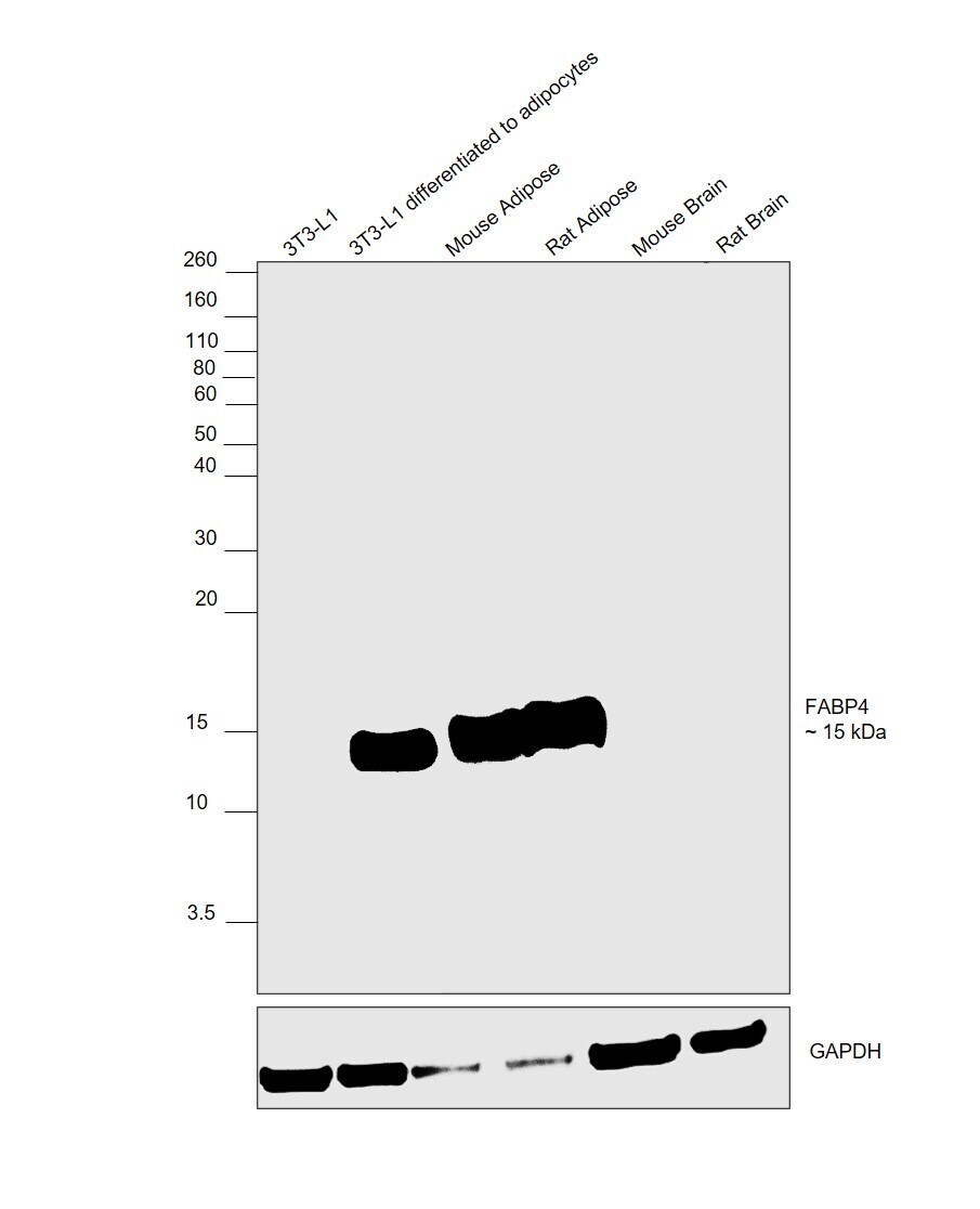
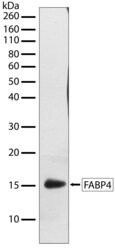
- Western blot was performed using Anti-FABP4 Recombinant Rabbit Monoclonal Antibody (2H3L2) (Product # 701158) and a 15 kDa band corresponding to Fatty acid-binding protein, adipocyte was observed across cell lines and tissue. Whole cell extracts (40 µg lysate) of 3T3-L1 (Lane 1), 3T3-L1 differentiated to adipocytes (Lane 2), Mouse Adipose (Lane 3), Rat Adipose (Lane 4), Mouse Brain (Lane 5), Rat Brain (Lane 6) were electrophoresed using NuPAGE™ 12% Bis-Tris Protein Gel (Product # NP0341BOX). Resolved proteins were then transferred onto a nitrocellulose membrane (Product # IB23001) by iBlot® 2 Dry Blotting System (Product # IB21001). The blot was probed with the primary antibody (0.5 µg/mL) and detected by chemiluminescence with Goat anti-Rabbit IgG (H+L) Superclonal™ Recombinant Secondary Antibody, HRP (Product # A27036,1:20,000 dilution using the iBright™ FL1500 Imaging System (Product # A44115). Chemiluminescent detection was performed using SuperSignal™ West Pico PLUS Chemiluminescent Substrate (Product # 34580). Upon differentiation of 3T3-L1 cells to adipocytes the expression of FABP4 should upregulate as observed. Also, a relative expression was observed in Adipose (high expressing tissue model) versus brain (low expressing tissue model) as reported.
- Submitted by
- Invitrogen Antibodies (provider)
- Main image
- Experimental details
- Western blot analysis of FABP4 in whole cell extracts of serum-starved 3T3 L1 cells treated with Insulin (100 ng/mL, 15 min) using a FABP4 recombinant rabbit monoclonal antibody (Product # 701158) at a dilution of 1 µg/mL. Samples were detected using chemiluminescence (ECL). Results show a band at ~15 kDa.

Supportive validation
- Submitted by
- Invitrogen Antibodies (provider)
- Main image
- Experimental details
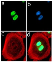
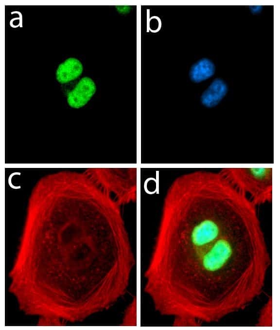
- Immunofluorescent analysis of FABP4 in HeLa cells using a FABP4 recombinant rabbit monoclonal antibody (Product # 701158) followed by detection using an Alexa Fluor 488-conjugated goat anti-rabbit secondary antibody (green) (Image A). Nuclei were stained using DAPI (Image B) and actin stained with Alexa Fluor 594 phalloidin (red) (image C). Image D is a composite image showing nuclear localization of FABP4.
Supportive validation
- Submitted by
- Invitrogen Antibodies (provider)
- Main image

- Experimental details
- Flow Cytometry analysis of FABP4 Receptor was done on 3T3-L1 cells (untreated, red histogram) and 3T3-L1 cells differentiation with adiponectin for 72 hours (blue histogram). Cells were fixed with 70% ethanol for 10 minutes, permeabilized with 0.25% Triton™ X-100 for 20 minutes, and blocked with 5% BSA for 30 minutes at room temperature. Cells were labeled with FABP4 Antibody, ABfinity™ Rabbit Monoclonal Antibody (Product # 701158) or with rabbit isotype control (pink histogram) at 3-5 µg/million cells in 2.5% BSA. After incubation at room temperature for 2 hours, the cells were labeled with Alexa Fluor® 488 Goat Anti-Rabbit Secondary Antibody (Product # A11008) at a dilution of 1:400 for 30 minutes at room temperature. The representative 10,000 cells were acquired and analyzed for each sample using an Attune® Acoustic Focusing Cytometer. The purple histogram represents unstained control cells.
