Explore
 Validate
 Learn
Learn Western blot
Antibody data
- Antibody Data
- Antigen structure
- References [11]
- Comments [0]
- Validations
- Western blot [2]
- Immunocytochemistry [2]
- Immunohistochemistry [1]
Submit
Validation data
Reference
Comment
Report error
- Product number
- AF1443 - Provider product page

- Provider
- R&D Systems
- Product name
- Mouse/Rat FABP4/A-FABP Antibody
- Antibody type
- Polyclonal
- Description
- Antigen Affinity-purified. Detects mouse FABP4 in direct ELISAs and Western blots. In direct ELISAs, less than 5% cross-reactivity with recombinant mouse (rm) FABP5 and rmFABP9 is observed.
- Reactivity
- Mouse, Rat
- Host
- Goat
- Conjugate
- Unconjugated
- Antigen sequence
P04117- Isotype
- IgG
- Vial size
- 100 ug
- Concentration
- LYOPH
- Storage
- Use a manual defrost freezer and avoid repeated freeze-thaw cycles. 12 months from date of receipt, -20 to -70 °C as supplied. 1 month, 2 to 8 °C under sterile conditions after reconstitution. 6 months, -20 to -70 °C under sterile conditions after reconstitution.
Submitted references Comparison of Immunosuppressive and Angiogenic Properties of Human Amnion-Derived Mesenchymal Stem Cells between 2D and 3D Culture Systems.
Maintenance of the bladder cancer precursor urothelial hyperplasia requires FOXA1 and persistent expression of oncogenic HRAS.
Single-cell analysis reveals fibroblast heterogeneity and myeloid-derived adipocyte progenitors in murine skin wounds.
A-FABP mediates adaptive thermogenesis by promoting intracellular activation of thyroid hormones in brown adipocytes.
Chemotherapy can induce weight normalization of morbidly obese mice despite undiminished ingestion of high fat diet.
Epigenetic programming of Dnmt3a mediated by AP2α is required for granting preadipocyte the ability to differentiate.
Epidermal Wnt/β-catenin signaling regulates adipocyte differentiation via secretion of adipogenic factors.
Artificial sweeteners stimulate adipogenesis and suppress lipolysis independently of sweet taste receptors.
Distinct fibroblast lineages determine dermal architecture in skin development and repair.
Age-related changes in rat bone-marrow mesenchymal stem cell plasticity.
Cyclooxygenase-2 deficiency attenuates adipose tissue differentiation and inflammation in mice.
Miceli V, Pampalone M, Vella S, Carreca AP, Amico G, Conaldi PG
Stem cells international 2019;2019:7486279
Stem cells international 2019;2019:7486279
Maintenance of the bladder cancer precursor urothelial hyperplasia requires FOXA1 and persistent expression of oncogenic HRAS.
Yee CH, Zheng Z, Shuman L, Yamashita H, Warrick JI, Wu XR, Raman JD, DeGraff DJ
Scientific reports 2019 Jan 22;9(1):270
Scientific reports 2019 Jan 22;9(1):270
Single-cell analysis reveals fibroblast heterogeneity and myeloid-derived adipocyte progenitors in murine skin wounds.
Guerrero-Juarez CF, Dedhia PH, Jin S, Ruiz-Vega R, Ma D, Liu Y, Yamaga K, Shestova O, Gay DL, Yang Z, Kessenbrock K, Nie Q, Pear WS, Cotsarelis G, Plikus MV
Nature communications 2019 Feb 8;10(1):650
Nature communications 2019 Feb 8;10(1):650
A-FABP mediates adaptive thermogenesis by promoting intracellular activation of thyroid hormones in brown adipocytes.
Shu L, Hoo RL, Wu X, Pan Y, Lee IP, Cheong LY, Bornstein SR, Rong X, Guo J, Xu A
Nature communications 2017 Jan 27;8:14147
Nature communications 2017 Jan 27;8:14147
Chemotherapy can induce weight normalization of morbidly obese mice despite undiminished ingestion of high fat diet.
Myers CE, Hoelzinger DB, Truong TN, Chew LA, Myles A, Chaudhuri L, Egan JB, Liu J, Gendler SJ, Cohen PA
Oncotarget 2017 Jan 17;8(3):5426-5438
Oncotarget 2017 Jan 17;8(3):5426-5438
Epigenetic programming of Dnmt3a mediated by AP2α is required for granting preadipocyte the ability to differentiate.
Guo W, Chen J, Yang Y, Zhu J, Wu J
Cell death & disease 2016 Dec 1;7(12):e2496
Cell death & disease 2016 Dec 1;7(12):e2496
Epidermal Wnt/β-catenin signaling regulates adipocyte differentiation via secretion of adipogenic factors.
Donati G, Proserpio V, Lichtenberger BM, Natsuga K, Sinclair R, Fujiwara H, Watt FM
Proceedings of the National Academy of Sciences of the United States of America 2014 Apr 15;111(15):E1501-9
Proceedings of the National Academy of Sciences of the United States of America 2014 Apr 15;111(15):E1501-9
Artificial sweeteners stimulate adipogenesis and suppress lipolysis independently of sweet taste receptors.
Simon BR, Parlee SD, Learman BS, Mori H, Scheller EL, Cawthorn WP, Ning X, Gallagher K, Tyrberg B, Assadi-Porter FM, Evans CR, MacDougald OA
The Journal of biological chemistry 2013 Nov 8;288(45):32475-89
The Journal of biological chemistry 2013 Nov 8;288(45):32475-89
Distinct fibroblast lineages determine dermal architecture in skin development and repair.
Driskell RR, Lichtenberger BM, Hoste E, Kretzschmar K, Simons BD, Charalambous M, Ferron SR, Herault Y, Pavlovic G, Ferguson-Smith AC, Watt FM
Nature 2013 Dec 12;504(7479):277-281
Nature 2013 Dec 12;504(7479):277-281
Age-related changes in rat bone-marrow mesenchymal stem cell plasticity.
Asumda FZ, Chase PB
BMC cell biology 2011 Oct 12;12:44
BMC cell biology 2011 Oct 12;12:44
Cyclooxygenase-2 deficiency attenuates adipose tissue differentiation and inflammation in mice.
Ghoshal S, Trivedi DB, Graf GA, Loftin CD
The Journal of biological chemistry 2011 Jan 7;286(1):889-98
The Journal of biological chemistry 2011 Jan 7;286(1):889-98
No comments: Submit comment
Supportive validation
- Submitted by
- R&D Systems (provider)
- Main image
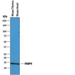
- Experimental details
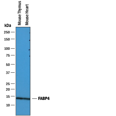
- Detection of Mouse FABP4/A-FABP by Western Blot. Western blot shows lysates of mouse thymus tissue and mouse heart tissue. PVDF membrane was probed with 0.25 µg/mL of Goat Anti-Mouse/Rat FABP4/A-FABP Antigen Affinity-purified Polyclonal Antibody (Catalog # AF1443) followed by HRP-conjugated Anti-Goat IgG Secondary Antibody (Catalog # HAF019). A specific band was detected for FABP4/A-FABP at approximately 14 kDa (as indicated). This experiment was conducted under reducing conditions and using Immunoblot Buffer Group 1.
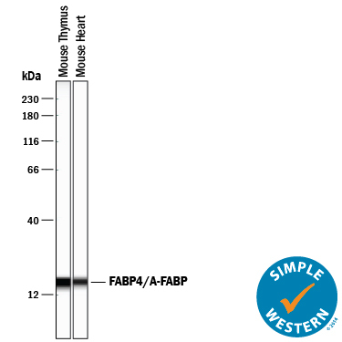
- Submitted by
- R&D Systems (provider)
- Main image

- Experimental details
- Detection of Mouse FABP4/A-FABP by Simple WesternTM. Simple Western lane view shows lysates of mouse thymus tissue and mouse heart tissue, loaded at 0.2 mg/mL. A specific band was detected for FABP4/A-FABP at approximately 17 kDa (as indicated) using 2.5 µg/mL of Goat Anti-Mouse/Rat FABP4/A-FABP Antigen Affinity-purified Polyclonal Antibody (Catalog # AF1443) followed by 1:50 dilution of HRP-conjugated Anti-Goat IgG Secondary Antibody (Catalog # HAF109). This experiment was conducted under reducing conditions and using the 12-230 kDa separation system.
Supportive validation
- Submitted by
- R&D Systems (provider)
- Main image

- Experimental details
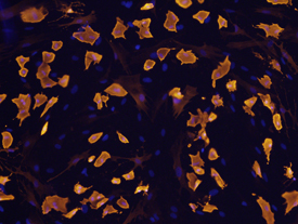
- FABP4 in Rat Mesenchymal Stem Cells. FABP4 was detected in immersion fixed rat mesenchymal stem cells differentiated to adipocytes using Goat Anti-Mouse/Rat FABP4 Antigen Affinity-purified Polyclonal Antibody (Catalog # AF1443) at 10 µg/mL for 3 hours at room temperature. Cells were stained using the NorthernLights™ 557-conjugated Anti-Goat IgG Secondary Antibody (yellow; Catalog # NL001) and counterstained with DAPI (blue). View our protocol for Fluorescent ICC Staining of Cells on Coverslips.
- Submitted by
- R&D Systems (provider)
- Main image

- Experimental details
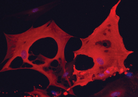
- FABP4 in Mouse Adipocytes. FABP4 was detected in immersion fixed mouse mesenchymal stem cell-derived adipocytes using 10 µg/mL Goat Anti-Mouse/Rat FABP4 Antigen Affinity-purified Polyclonal Antibody (Catalog # AF1443) for 3 hours at room temperature. Cells were stained with the NorthernLights™ 557-conjugated Anti-Goat IgG Secondary Antibody (red; Catalog # NL001) and counterstained with DAPI (blue). View our protocol for Fluorescent ICC Staining of Cells on Coverslips.
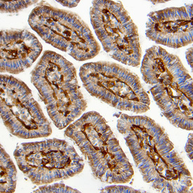
Supportive validation
- Submitted by
- R&D Systems (provider)
- Main image
- Experimental details
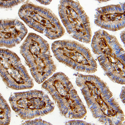
- FABP4 in Mouse Intestine. FABP4 was detected in perfusion fixed frozen sections of mouse intestine using Goat Anti-Mouse/Rat FABP4 Antigen Affinity-purified Polyclonal Antibody (Catalog # AF1443) at 5 µg/mL overnight at 4 °C. Tissue was stained using the Anti-Goat HRP-DAB Cell & Tissue Staining Kit (brown; Catalog # CTS008) and counterstained with hematoxylin (blue). Specific labeling was localized to the plasma membrane of epithelial cells in intestinal glands. View our protocol for Chromogenic IHC Staining of Frozen Tissue Sections.