 Explore
 Validate
 Learn
Learn Western blot
 ELISA
ELISA Immunocytochemistry
Antibody data
- Antibody Data
- Antigen structure
- References [6]
- Comments [0]
- Validations
- Immunocytochemistry [2]
- Immunohistochemistry [1]
- Other assay [4]
Submit
Validation data
Reference
Comment
Report error
- Product number
- PA5-30591 - Provider product page

- Provider
- Invitrogen Antibodies
- Product name
- FABP4 Polyclonal Antibody
- Antibody type
- Polyclonal
- Antigen
- Synthetic peptide
- Description
- Recommended positive controls: 293T, A431, HeLa, HepG2. Predicted reactivity: Rhesus Monkey (100%). Store product as a concentrated solution. Centrifuge briefly prior to opening the vial.
- Reactivity
- Human, Mouse
- Host
- Rabbit
- Isotype
- IgG
- Vial size
- 100 μL
- Concentration
- 0.19 mg/mL
- Storage
- Store at 4°C short term. For long term storage, store at -20°C, avoiding freeze/thaw cycles.
Submitted references Immunity-and-matrix-regulatory cells derived from human embryonic stem cells safely and effectively treat mouse lung injury and fibrosis.
In vitro osteoblastic differentiation of mesenchymal stem cells generates cell layers with distinct properties.
A fully defined static suspension culture system for large-scale human embryonic stem cell production.
Heterogeneous Differentiation of Human Mesenchymal Stem Cells in 3D Extracellular Matrix Composites.
Tube formation in the first trimester placental trophoblast cells: Differential effects of angiogenic growth factors and fatty acids.
Age-dependent changes in TDP-43 levels in a mouse model of Alzheimer disease are linked to Aβ oligomers accumulation.
Wu J, Song D, Li Z, Guo B, Xiao Y, Liu W, Liang L, Feng C, Gao T, Chen Y, Li Y, Wang Z, Wen J, Yang S, Liu P, Wang L, Wang Y, Peng L, Stacey GN, Hu Z, Feng G, Li W, Huo Y, Jin R, Shyh-Chang N, Zhou Q, Wang L, Hu B, Dai H, Hao J
Cell research 2020 Sep;30(9):794-809
Cell research 2020 Sep;30(9):794-809
In vitro osteoblastic differentiation of mesenchymal stem cells generates cell layers with distinct properties.
Hanna H, Mir LM, Andre FM
Stem cell research & therapy 2018 Jul 27;9(1):203
Stem cell research & therapy 2018 Jul 27;9(1):203
A fully defined static suspension culture system for large-scale human embryonic stem cell production.
Li X, Ma R, Gu Q, Liang L, Wang L, Zhang Y, Wang X, Liu X, Li Z, Fang J, Wu J, Wang Y, Li W, Hu B, Wang L, Zhou Q, Hao J
Cell death & disease 2018 Aug 30;9(9):892
Cell death & disease 2018 Aug 30;9(9):892
Heterogeneous Differentiation of Human Mesenchymal Stem Cells in 3D Extracellular Matrix Composites.
Jung JP, Bache-Wiig MK, Provenzano PP, Ogle BM
BioResearch open access 2016;5(1):37-48
BioResearch open access 2016;5(1):37-48
Tube formation in the first trimester placental trophoblast cells: Differential effects of angiogenic growth factors and fatty acids.
Pandya AD, Das MK, Sarkar A, Vilasagaram S, Basak S, Duttaroy AK
Cell biology international 2016 Jun;40(6):652-61
Cell biology international 2016 Jun;40(6):652-61
Age-dependent changes in TDP-43 levels in a mouse model of Alzheimer disease are linked to Aβ oligomers accumulation.
Caccamo A, Magrí A, Oddo S
Molecular neurodegeneration 2010 Nov 11;5:51
Molecular neurodegeneration 2010 Nov 11;5:51
No comments: Submit comment
Supportive validation
- Submitted by
- Invitrogen Antibodies (provider)
- Main image
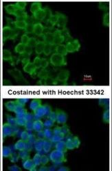
- Experimental details
- Immunofluorescent analysis of FABP4 in paraformaldehyde-fixed mouse ESC D3 cells using a FABP4 polyclonal antibody (Product # PA5-30591) at a 1:200 dilution.
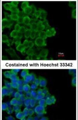
- Submitted by
- Invitrogen Antibodies (provider)
- Main image
- Experimental details
- Immunofluorescent analysis of FABP4 in paraformaldehyde-fixed HeLa cells using a FABP4 polyclonal antibody (Product # PA5-30591) at a 1:200 dilution.

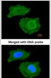
Supportive validation
- Submitted by
- Invitrogen Antibodies (provider)
- Main image
- Experimental details
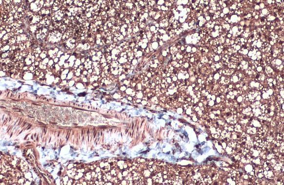
- FABP4 Polyclonal Antibody detects FABP4 protein at cytoplasm and nucleus by immunohistochemical analysis. Sample: Paraffin-embedded mouse brown adipocyte. FABP4 stained by FABP4 Polyclonal Antibody (Product # PA5-30591) diluted at 1:500. Antigen Retrieval: Citrate buffer, pH 6.0, 15 min.
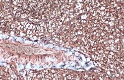
Supportive validation
- Submitted by
- Invitrogen Antibodies (provider)
- Main image
- Experimental details
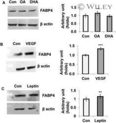
- NULL

- Submitted by
- Invitrogen Antibodies (provider)
- Main image
- Experimental details
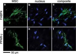
- FIG. 4. Expression of FABP4 and PPAR-gamma proteins in 3D ColI composites after 28 days of culture. Immunofluorescence was visualized by MPLSM. FABP4 (a-c) and PPAR-gamma (d-f) . Adipogenic protein markers (green) and nuclei (blue); scale bar 50 mum.

- Submitted by
- Invitrogen Antibodies (provider)
- Main image
- Experimental details
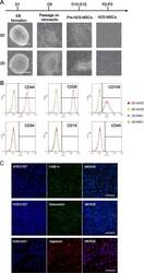
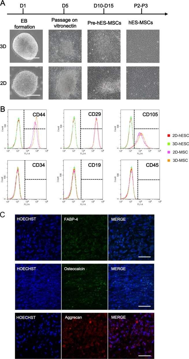
- Fig. 4 Comparison of MSCs derived from 2D-hESCs and 3D-hESCs. a The morphology of 2D- and 3D-hESC-MSCs at different stages of differentiation. b Flow cytometry analysis revealed specific MSC surface markers (CD44, CD29, and CD105) with negative controls (CD34, CD19, and CD45) in 2D- and 3D-hESC-MSCs. c Immunostaining of differentiated 3D-hESC-MSCs expressing an adipocyte marker (FABP-4), osteocytes maker (osteocalcin), and chondrocytes marker (aggrecan)
- Submitted by
- Invitrogen Antibodies (provider)
- Main image
- Experimental details
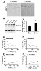
- Figure 4 TDP-43 and TDP-35 levels do not correlate with tau phosphorylated at Thr181 . (A) Representative microphotographs of 3xTg-AD brain sections depicting hippocampal CA1 pyramidal neurons stained with the anti-tau antibody AT270, which recognize tau phosphorylated at Thr181 (n = 6/age group). (B) Representative Western blots of proteins extracted from 6- and 12-month old 3xTg-AD mice probed with the AT270 antibody. beta-actin is used as a loading control. (C) Quantitative analysis of the blots shows that the steady-state levels of AT270 were significantly higher in the brains of 12-month-old 3xTg-AD mice compared to 6-month-old mice (n = 6/age group; p < 0.05). Data are presented as +- SEM and analyzed by t-test analysis. (D-E) Linear regression analysis indicated that the levels of AT270 in the brains of 6-month-old 3xTg-AD mice did not correlate with the levels of TDP-43 (r 2 = 0.09148, p = 0.56) and TDP-35 (r 2 = 0.1491, p = 0.45). (F-G) Similarly, no correlation between AT270 and TDP-43 (r 2 = 0.4644, p = 0.14) and TDP-35 (r 2 = 0.4469, p = 0.15) levels was observed at 12 months of age.
