Explore
 Validate
 Learn
Learn Western blot
 Immunocytochemistry
Antibody data
- Antibody Data
- Antigen structure
- References [2]
- Comments [0]
- Validations
- Immunocytochemistry [1]
Submit
Validation data
Reference
Comment
Report error
- Product number
- MAB2066 - Provider product page

- Provider
- R&D Systems
- Product name
- Human/Rat/Chicken Laminin S Antibody
- Antibody type
- Monoclonal
- Description
- Protein A or G purified from hybridoma culture supernatant. Detects human, rat, and chicken Laminin S in Western blots.
- Reactivity
- Human, Rat, Chicken/Avian
- Host
- Mouse
- Conjugate
- Unconjugated
- Isotype
- IgG
- Antibody clone number
- C4
- Vial size
- 100 ug
- Concentration
- LYOPH
- Storage
- Use a manual defrost freezer and avoid repeated freeze-thaw cycles. 12 months from date of receipt, -20 to -70 °C as supplied. 1 month, 2 to 8 °C under sterile conditions after reconstitution. 6 months, -20 to -70 °C under sterile conditions after reconstitution.
Submitted references Bioengineering Human Neurological Constructs Using Decellularized Meningeal Scaffolds for Application in Spinal Cord Injury.
Synthesis and assembly of the synaptic cleft protein S-laminin by cultured cells.
Vishwakarma SK, Bardia A, Lakkireddy C, Paspala SAB, Khan AA
Frontiers in bioengineering and biotechnology 2018;6:150
Frontiers in bioengineering and biotechnology 2018;6:150
Synthesis and assembly of the synaptic cleft protein S-laminin by cultured cells.
Green TL, Hunter DD, Chan W, Merlie JP, Sanes JR
The Journal of biological chemistry 1992 Jan 25;267(3):2014-22
The Journal of biological chemistry 1992 Jan 25;267(3):2014-22
No comments: Submit comment
Supportive validation
- Submitted by
- R&D Systems (provider)
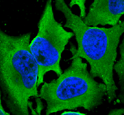
- Main image

- Experimental details
- Laminin S in HeLa Human Cell Line. Laminin S was detected in immersion fixed HeLa human cervical epithelial carcinoma cell line using Mouse Anti-Human/Rat/Chicken Laminin S Monoclonal Antibody (Catalog # MAB2066) at 10 µg/mL for 3 hours at room temperature. Cells were stained using the NorthernLights™ 493-conjugated Anti-Mouse IgG Secondary Antibody (green; Catalog # NL009) and counterstained with DAPI (blue). Specific staining was localized to cytoplasm. View our protocol for Fluorescent ICC Staining of Cells on Coverslips.