 Explore
 Validate
 Learn
Learn Western blot
Antibody data
- Antibody Data
- Antigen structure
- References [3]
- Comments [0]
- Validations
- Western blot [2]
- Immunohistochemistry [1]
Submit
Validation data
Reference
Comment
Report error
- Product number
- AF3074 - Provider product page

- Provider
- R&D Systems
- Product name
- Human Thrombospondin-1 Antibody
- Antibody type
- Polyclonal
- Description
- Antigen Affinity-purified. Detects human Thrombospondin-1 in direct ELISAs and Western blots. In direct ELISAs, less than 5% cross-reactivity with recombinant human (rh) Thrombospondin-2 and rhThrombospondin-4 is observed.
- Reactivity
- Human
- Host
- Goat
- Conjugate
- Unconjugated
- Antigen sequence
CAA32889- Isotype
- IgG
- Vial size
- 100 ug
- Concentration
- LYOPH
- Storage
- Use a manual defrost freezer and avoid repeated freeze-thaw cycles. 12 months from date of receipt, -20 to -70 °C as supplied. 1 month, 2 to 8 °C under sterile conditions after reconstitution. 6 months, -20 to -70 °C under sterile conditions after reconstitution.
Submitted references Subretinal Human Umbilical Tissue-Derived Cell Transplantation Preserves Retinal Synaptic Connectivity and Attenuates Müller Glial Reactivity.
Thrombin-reduced miR-27b attenuates platelet angiogenic activities in vitro via enhancing platelet synthesis of anti-angiogenic thrombospondin-1.
Canonical wnt signaling regulates extracellular matrix expression in the trabecular meshwork.
Koh S, Chen WJ, Dejneka NS, Harris IR, Lu B, Girman S, Saylor J, Wang S, Eroglu C
The Journal of neuroscience : the official journal of the Society for Neuroscience 2018 Mar 21;38(12):2923-2943
The Journal of neuroscience : the official journal of the Society for Neuroscience 2018 Mar 21;38(12):2923-2943
Thrombin-reduced miR-27b attenuates platelet angiogenic activities in vitro via enhancing platelet synthesis of anti-angiogenic thrombospondin-1.
Miao X, Rahman MF, Jiang L, Min Y, Tan S, Xie H, Lee L, Wang M, Malmström RE, Lui WO, Li N
Journal of thrombosis and haemostasis : JTH 2018 Apr;16(4):791-801
Journal of thrombosis and haemostasis : JTH 2018 Apr;16(4):791-801
Canonical wnt signaling regulates extracellular matrix expression in the trabecular meshwork.
Villarreal G Jr, Chatterjee A, Oh SS, Oh DJ, Kang MH, Rhee DJ
Investigative ophthalmology & visual science 2014 Oct 28;55(11):7433-40
Investigative ophthalmology & visual science 2014 Oct 28;55(11):7433-40
No comments: Submit comment
Supportive validation
- Submitted by
- R&D Systems (provider)
- Main image
- Experimental details
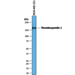
- Detection of Human Thrombospondin-1 by Western Blot. Western blot shows lysates of MDA-MB-231 human breast cancer cell line. PVDF membrane was probed with 1 µg/mL of Goat Anti-Human Thrombospondin-1 Antigen Affinity-purified Polyclonal Antibody (Catalog # AF3074) followed by HRP-conjugated Anti-Goat IgG Secondary Antibody (Catalog # HAF019). A specific band was detected for Thrombospondin-1 at approximately 160 kDa (as indicated). This experiment was conducted under reducing conditions and using Immunoblot Buffer Group 1.

- Submitted by
- R&D Systems (provider)
- Main image
- Experimental details
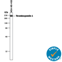
- Detection of Human Thrombospondin-1 by Simple WesternTM. Simple Western lane view shows lysates of MBA-MB-468 human breast cancer cell line, loaded at 0.2 mg/mL. A specific band was detected for Thrombospondin-1 at approximately 221 kDa (as indicated) using 10 µg/mL of Goat Anti-Human Thrombospondin-1 Antigen Affinity-purified Polyclonal Antibody (Catalog # AF3074) followed by 1:50 dilution of HRP-conjugated Anti-Goat IgG Secondary Antibody (Catalog # HAF109). This experiment was conducted under reducing conditions and using the 12-230 kDa separation system.

Supportive validation
- Submitted by
- R&D Systems (provider)
- Main image
- Experimental details
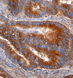
- Thrombospondin-1 in Human Colon. Thrombospondin-1 was detected in immersion fixed paraffin-embedded sections of human colon using Goat Anti-Human Thrombospondin-1 Antigen Affinity-purified Polyclonal Antibody (Catalog # AF3074) at 1.7 µg/mL overnight at 4 °C. Tissue was stained using the Anti-Goat HRP-DAB Cell & Tissue Staining Kit (brown; Catalog # CTS008) and counterstained with hematoxylin (blue). Specific staining was localized to the cytoplasm of epithelial cells. View our protocol for Chromogenic IHC Staining of Paraffin-embedded Tissue Sections.
