 Explore
 Validate
 Learn
Learn Western blot
Antibody data
- Antibody Data
- Antigen structure
- References [6]
- Comments [0]
- Validations
- Western blot [1]
- Immunohistochemistry [1]
Submit
Validation data
Reference
Comment
Report error
- Product number
- AF1334 - Provider product page

- Provider
- R&D Systems
- Product name
- Human IGFBP-rp1/IGFBP-7 Antibody
- Antibody type
- Polyclonal
- Description
- Antigen Affinity-purified. Detects human IGFBP-rp1 in direct ELISAs and Western blots. In direct ELISAs, less than 5% cross-reactivity with recombinant human (rh) IGFBP-L1, rhIGFBP-rp10 and recombinant mouse IGFBP-7 is observed.
- Reactivity
- Human
- Host
- Goat
- Conjugate
- Unconjugated
- Antigen sequence
AAA16187- Isotype
- IgG
- Vial size
- 100 ug
- Concentration
- LYOPH
- Storage
- Use a manual defrost freezer and avoid repeated freeze-thaw cycles. 12 months from date of receipt, -20 to -70 °C as supplied. 1 month, 2 to 8 °C under sterile conditions after reconstitution. 6 months, -20 to -70 °C under sterile conditions after reconstitution.
Submitted references IGFBP7, a novel tumor stroma marker, with growth-promoting effects in colon cancer through a paracrine tumor-stroma interaction.
Disulfiram suppresses growth of the malignant pleural mesothelioma cells in part by inducing apoptosis.
Regulation of human skin pigmentation in situ by repetitive UV exposure: molecular characterization of responses to UVA and/or UVB.
Glioblastoma-secreted factors induce IGFBP7 and angiogenesis by modulating Smad-2-dependent TGF-beta signaling.
Insulin-like growth factor binding protein 7 mediates glioma cell growth and migration.
Insulin-like growth factor binding protein 7 mediates glioma cell growth and migration.
Rupp C, Scherzer M, Rudisch A, Unger C, Haslinger C, Schweifer N, Artaker M, Nivarthi H, Moriggl R, Hengstschläger M, Kerjaschki D, Sommergruber W, Dolznig H, Garin-Chesa P
Oncogene 2015 Feb 12;34(7):815-25
Oncogene 2015 Feb 12;34(7):815-25
Disulfiram suppresses growth of the malignant pleural mesothelioma cells in part by inducing apoptosis.
Cheriyan VT, Wang Y, Muthu M, Jamal S, Chen D, Yang H, Polin LA, Tarca AL, Pass HI, Dou QP, Sharma S, Wali A, Rishi AK
PloS one 2014;9(4):e93711
PloS one 2014;9(4):e93711
Regulation of human skin pigmentation in situ by repetitive UV exposure: molecular characterization of responses to UVA and/or UVB.
Choi W, Miyamura Y, Wolber R, Smuda C, Reinhold W, Liu H, Kolbe L, Hearing VJ
The Journal of investigative dermatology 2010 Jun;130(6):1685-96
The Journal of investigative dermatology 2010 Jun;130(6):1685-96
Glioblastoma-secreted factors induce IGFBP7 and angiogenesis by modulating Smad-2-dependent TGF-beta signaling.
Pen A, Moreno MJ, Durocher Y, Deb-Rinker P, Stanimirovic DB
Oncogene 2008 Nov 20;27(54):6834-44
Oncogene 2008 Nov 20;27(54):6834-44
Insulin-like growth factor binding protein 7 mediates glioma cell growth and migration.
Jiang W, Xiang C, Cazacu S, Brodie C, Mikkelsen T
Neoplasia (New York, N.Y.) 2008 Dec;10(12):1335-42
Neoplasia (New York, N.Y.) 2008 Dec;10(12):1335-42
Insulin-like growth factor binding protein 7 mediates glioma cell growth and migration.
Jiang W, Xiang C, Cazacu S, Brodie C, Mikkelsen T
Neoplasia (New York, N.Y.) 2008 Dec;10(12):1335-42
Neoplasia (New York, N.Y.) 2008 Dec;10(12):1335-42
No comments: Submit comment
Supportive validation
- Submitted by
- R&D Systems (provider)
- Main image

- Experimental details
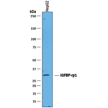
- Detection of Human IGFBP-rp1/IGFBP-7 by Western Blot. Western blot shows lysates of HepG2 human hepatocellular carcinoma cell line. PVDF membrane was probed with 1 µg/mL of Goat Anti-Human IGFBP-rp1/IGFBP-7 Antigen Affinity-purified Polyclonal Antibody (Catalog # AF1334) followed by HRP-conjugated Anti-Goat IgG Secondary Antibody (Catalog # HAF019). A specific band was detected for IGFBP-rp1/IGFBP-7 at approximately 30 kDa (as indicated). This experiment was conducted under reducing conditions and using Immunoblot Buffer Group 1.
Supportive validation
- Submitted by
- R&D Systems (provider)
- Main image
- Experimental details
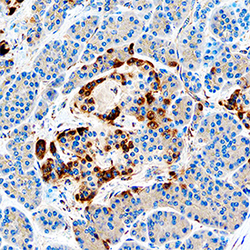
- IGFBP-rp1/IGFBP-7 in Human Pancreas. IGFBP-rp1/IGFBP-7 was detected in immersion fixed paraffin-embedded sections of human pancreas using Goat Anti-Human IGFBP-rp1/IGFBP-7 Antigen Affinity-purified Polyclonal Antibody (Catalog # AF1334) at 0.3 µg/mL overnight at 4 °C. Tissue was stained using the Anti-Goat HRP-DAB Cell & Tissue Staining Kit (brown; Catalog # CTS008) and counterstained with hematoxylin (blue). Specific staining was localized to islets. View our protocol for Chromogenic IHC Staining of Paraffin-embedded Tissue Sections.
