 Explore
 Validate
 Learn
Learn Western blot
 Immunocytochemistry
 Immunohistochemistry
Antibody data
- Antibody Data
- Antigen structure
- References [4]
- Comments [0]
- Validations
- Western blot [1]
- Immunocytochemistry [1]
Submit
Validation data
Reference
Comment
Report error
- Product number
- HPA017893 - Provider product page

- Provider
- Atlas Antibodies
- Proper citation
- Atlas Antibodies Cat#HPA017893, RRID:AB_1852274
- Product name
- Anti-RRP1B
- Antibody type
- Polyclonal
- Description
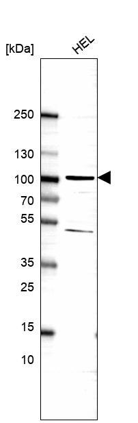
- Polyclonal Antibody against Human RRP1B, Gene description: ribosomal RNA processing 1B, Alternative Gene Names: KIAA0179, Nnp1, PPP1R136, RRP1, Validated applications: IHC, WB, ICC, Uniprot ID: Q14684, Storage: Store at +4°C for short term storage. Long time storage is recommended at -20°C.
- Reactivity
- Human
- Host
- Rabbit
- Conjugate
- Unconjugated
- Isotype
- IgG
- Vial size
- 100 µl
- Concentration
- 0.5 mg/ml
- Storage
- Store at +4°C for short term storage. Long time storage is recommended at -20°C.
- Handling
- The antibody solution should be gently mixed before use.
Submitted references
BRD4 Short Isoform Interacts with RRP1B, SIPA1 and Components of the LINC Complex at the Inner Face of the Nuclear Membrane
RRP1B is a metastasis modifier that regulates the expression of alternative mRNA isoforms through interactions with SRSF1
BRD4 Short Isoform Interacts with RRP1B, SIPA1 and Components of the LINC Complex at the Inner Face of the Nuclear Membrane
Quinodoz S, Jiang L, Abu-Alfa A, Comi T, Zhao H, Yu Q, Wiesner L, Botello J, Donlic A, Soehalim E, Zorbas C, Wacheul L, Košmrlj A, Lafontaine D, Klinge S, Brangwynne C
2024
2024
BRD4 Short Isoform Interacts with RRP1B, SIPA1 and Components of the LINC Complex at the Inner Face of the Nuclear Membrane
Samant R, Alsarraj J, Faraji F, Geiger T, Mattaini K, Williams M, Wu J, Ha N, Merlino T, Walker R, Bosley A, Xiao Z, Andresson T, Esposito D, Smithers N, Lugo D, Prinjha R, Day A, Crawford N, Ozato K, Gardner K, Hunter K
PLoS ONE 2013;8(11):e80746
PLoS ONE 2013;8(11):e80746
RRP1B is a metastasis modifier that regulates the expression of alternative mRNA isoforms through interactions with SRSF1
Lee M, Dworkin A, Gildea D, Trivedi N, Moorhead G, Crawford N
Oncogene 2013;33(14):1818-1827
Oncogene 2013;33(14):1818-1827
BRD4 Short Isoform Interacts with RRP1B, SIPA1 and Components of the LINC Complex at the Inner Face of the Nuclear Membrane
Alsarraj J, Faraji F, Geiger T, Mattaini K, Williams M, Wu J, Ha N, Merlino T, Walker R, Bosley A, Xiao Z, Andresson T, Esposito D, Smithers N, Lugo D, Prinjha R, Day A, Crawford N, Ozato K, Gardner K, Hunter K, Samant R
PLoS ONE 2013 November;8(11)
PLoS ONE 2013 November;8(11)
No comments: Submit comment
Enhanced validation
- Submitted by
- Atlas Antibodies (provider)
- Enhanced method
- Genetic validation
- Main image

- Experimental details
- Western blot analysis in U2OS cells transfected with control siRNA, target specific siRNA probe #1 and #2, using Anti-RRP1B antibody. Remaining relative intensity is presented.
- Sample type
- Human
- Protocol
- Protocol
Supportive validation
- Submitted by
- Atlas Antibodies (provider)
- Main image

- Experimental details
- Immunofluorescent staining of human cell line A-431 shows localization to nucleoli.
- Sample type
- Human
