Explore
 Validate
 Learn
LearnTA328665
antibody from OriGene
Targeting: AGTR1
AG2S, AGTR1A, AGTR1B, AT1, AT1B, AT2R1, AT2R1A, AT2R1B, HAT1R
 Western blot
Antibody data
- Antibody Data
- Antigen structure
- References [0]
- Comments [0]
- Validations
- Western blot [2]
- Immunocytochemistry [1]
- Immunohistochemistry [1]
- Flow cytometry [1]
Submit
Validation data
Reference
Comment
Report error
- Product number
- TA328665 - Provider product page

- Provider
- OriGene
- Product name
- Rabbit Polyclonal Anti-Angiotensin II Receptor Type-1 (extracellular)
- Antibody type
- Polyclonal
- Description
- Rabbit Polyclonal Anti-Angiotensin II Receptor Type-1 (extracellular)
- Host
- Rabbit
- Conjugate
- Unconjugated
- Epitope
- AGTR1
- Antibody clone number
- NULL
- Vial size
- 200 µl
- Concentration
- NULL
No comments: Submit comment
Supportive validation
- Submitted by
- OriGene (provider)
- Main image
- Experimental details
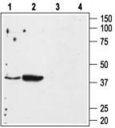
- Western blot analysis of mouse kidney (lanes 1 and 3) and mouse heart (lanes 2 and 4) membranes: 1, 2. Anti-Angiotensin II Receptor Type-1 (extracellular) antibody, (1:500). 3, 4. Anti-Angiotensin II Receptor Type-1 (extracellular) antibody, preincubated with the control peptide antigen.
- Validation comment
- WB
- Submitted by
- OriGene (provider)
- Main image
- Experimental details
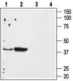
- Western blot analysis of rat liver (lanes 1 and 3) and rat kidney (lanes 2 and 4) membranes: 1, 2. Anti-Angiotensin II Receptor Type-1 (extracellular) antibody, (1:200). 3, 4. Anti-Angiotensin II Receptor Type-1 (extracellular) antibody, preincubated with the control peptide antigen.
- Validation comment
- WB

Supportive validation
- Submitted by
- OriGene (provider)
- Main image
- Experimental details
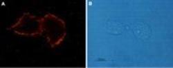
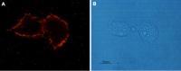
- Expression of Angiotensin II Receptor Type-1 in rat C6 glioma cells. Immunocytochemical staining of live intact rat C6 glioma cells. A. Cells were stained using Anti-Angiotensin II Receptor Type-1 (extracellular) antibody, (1:100), followed by goat anti-rabbit-AlexaFluor-555 secondary antibody. B. Live intact C6 cells.
- Validation comment
- IF
Supportive validation
- Submitted by
- OriGene (provider)
- Main image
- Experimental details
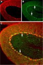
- Expression of Angiotensin II Receptor Type-1 in mouse cerebellum. Immunohistochemical staining of mouse cerebellum using Anti-Angiotensin II Receptor Type-1 (extracellular) antibody. A. Mouse anti-Parvalbumin (red) is detected in the Purkinje layer. B. In the same section, AT1 receptor (green) is also present in the Purkinje layer. Arrows point at AT1 receptor immunoreactive cells. Merge of A and B pannels reveals partial co-localization.
- Validation comment
- IHC

Supportive validation
- Submitted by
- OriGene (provider)
- Main image

- Experimental details
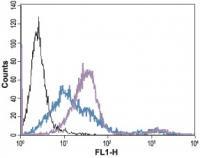
- Indirect flow cytometry analysis of live intact human Jurkat T-cell leukemia cells: black line, Unstained cells + goat-anti-rabbit-FITC. blue line, Cells + Anti-Angiotensin II Receptor Type-1 (extracellular) antibody, (5 ug) + goat-anti-rabbit-FITC. purple line, Cells + Anti-Angiotensin II Receptor Type-1 (extracellular) antibody, (10 ug) + goat-anti-rabbit-FITC.
- Validation comment
- FC