Explore
 Validate
 Learn
LearnNB100-57073
antibody from Novus Biologicals
Targeting: AGTR1
AG2S, AGTR1A, AGTR1B, AT1, AT1B, AT2R1, AT2R1A, AT2R1B, HAT1R
 Western blot
 ELISA
ELISAAntibody data
- Antibody Data
- Antigen structure
- References [2]
- Comments [0]
- Validations
- Western blot [2]
Submit
Validation data
Reference
Comment
Report error
- Product number
- NB100-57073 - Provider product page

- Provider
- Novus Biologicals
- Proper citation
- Novus Cat#NB100-57073, RRID:AB_2225562
- Product name
- Goat Polyclonal AGTR-1 Antibody
- Antibody type
- Polyclonal
- Description
- Immunogen affinity purified. All reported variants represent identical protein (NP_000676.1, NP_004826.2, NP_033611.1, NP_114038.1 and NP_114438.1).
- Reactivity
- Human, Mouse
- Host
- Goat
- Isotype
- IgG
- Vial size
- 0.1 mg
- Concentration
- 0.5 mg/ml
- Storage
- Store at -20C. Avoid freeze-thaw cycles.
Submitted references Amelioration of systemic fibrosis in mice by angiotensin II receptor blockade.
Role of brain angiotensin AT1 receptor in the carbachol-induced natriuresis and expression of nNOS in the locus coeruleus and proximal convoluted tubule.
Marut W, Kavian N, Servettaz A, Hua-Huy T, Nicco C, Chéreau C, Weill B, Dinh-Xuan AT, Batteux F
Arthritis and rheumatism 2013 May;65(5):1367-77
Arthritis and rheumatism 2013 May;65(5):1367-77
Role of brain angiotensin AT1 receptor in the carbachol-induced natriuresis and expression of nNOS in the locus coeruleus and proximal convoluted tubule.
Wang M, Jiang CL, Wang CY, Yao QY
Physiological research 2007;56(4):383-91
Physiological research 2007;56(4):383-91
No comments: Submit comment
Supportive validation
- Submitted by
- Novus Biologicals (provider)
- Main image

- Experimental details
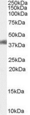
- Western Blot: AGTR-1 Antibody [NB100-57073] - Staining of Mouse Liver lysate (35 ug protein in RIPA buffer). Primary incubation was 1 hour. Detected by chemiluminescence
- Submitted by
- Novus Biologicals (provider)
- Main image

- Experimental details
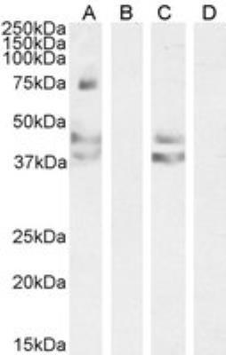
- Western Blot: AGTR-1 Antibody [NB100-57073] - Staining of Human Liver (A) + peptide (B), Mouse Liver (C) + peptide (D) lysate (35 ug protein in RIPA buffer). Antibody at 0.2 ug/mL. Detected by chemiluminescence.