Explore
 Validate
 Learn
Learn Western blot
 Immunocytochemistry
Antibody data
- Antibody Data
- Antigen structure
- References [5]
- Comments [0]
- Validations
- Immunocytochemistry [1]
- Immunohistochemistry [1]
Submit
Validation data
Reference
Comment
Report error
- Product number
- PA3-067 - Provider product page

- Provider
- Invitrogen Antibodies
- Product name
- Endothelin 1 Polyclonal Antibody
- Antibody type
- Polyclonal
- Antigen
- Other
- Description
- PA3-067 detects Endothelin 1 from human, mouse, and rat samples. PA3-067 has been successfully used in Western blot, immunocytochemistry, and immunohistochemistry procedures. By Western blot, this antibody detects a 24.4 kDa protein corresponding to mouse Endothelin 1. The PA3-067 antigen is mouse Endothelin-1 CS-Abu-SSLMDKE-Abu-VYF-Abu-HLDIIW coupled to KLH.
- Reactivity
- Human, Mouse, Rat
- Host
- Rabbit
- Isotype
- IgG
- Vial size
- 100 μL
- Concentration
- Conc. Not Determined
- Storage
- -20°C, Avoid Freeze/Thaw Cycles
Submitted references Evaluation of Neuroprotective Effect of Sevoflurane in Acute Traumatic Brain Injury: An Experimental Study in Rats.
N-methyl-D-aspartate Receptor Antagonists may Ameliorate Spinal Cord Injury by Inhibiting Oxidative Stress: An Experimental Study in Rats.
Activated Endothelial TGFβ1 Signaling Promotes Venous Thrombus Nonresolution in Mice Via Endothelin-1: Potential Role for Chronic Thromboembolic Pulmonary Hypertension.
Expression of vascular endothelial growth factor and glial fibrillary acidic protein in a rat model of traumatic brain injury treated with honokiol: a biochemical and immunohistochemical study.
Histopathological changes in the choroid plexus after traumatic brain injury in the rats: a histologic and immunohistochemical study.
Dogan G, Karaca O
Turkish neurosurgery 2020;30(2):237-243
Turkish neurosurgery 2020;30(2):237-243
N-methyl-D-aspartate Receptor Antagonists may Ameliorate Spinal Cord Injury by Inhibiting Oxidative Stress: An Experimental Study in Rats.
Dogan G, Karaca O
Turkish neurosurgery 2020;30(1):60-68
Turkish neurosurgery 2020;30(1):60-68
Activated Endothelial TGFβ1 Signaling Promotes Venous Thrombus Nonresolution in Mice Via Endothelin-1: Potential Role for Chronic Thromboembolic Pulmonary Hypertension.
Bochenek ML, Leidinger C, Rosinus NS, Gogiraju R, Guth S, Hobohm L, Jurk K, Mayer E, Münzel T, Lankeit M, Bosmann M, Konstantinides S, Schäfer K
Circulation research 2020 Jan 17;126(2):162-181
Circulation research 2020 Jan 17;126(2):162-181
Expression of vascular endothelial growth factor and glial fibrillary acidic protein in a rat model of traumatic brain injury treated with honokiol: a biochemical and immunohistochemical study.
Çetin A, Deveci E
Folia morphologica 2019;78(4):684-694
Folia morphologica 2019;78(4):684-694
Histopathological changes in the choroid plexus after traumatic brain injury in the rats: a histologic and immunohistochemical study.
Özevren H, Deveci E, Tuncer MC
Folia morphologica 2018;77(4):642-648
Folia morphologica 2018;77(4):642-648
No comments: Submit comment
Supportive validation
- Submitted by
- Invitrogen Antibodies (provider)
- Main image
- Experimental details
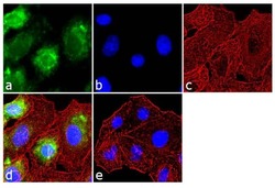
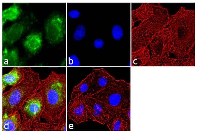
- Immunofluorescence analysis of Endothelin 1 was performed using 70% confluent log phase A549 cells. The cells were fixed with 4% paraformaldehyde for 10 minutes, permeabilized with 0.1% Triton™ X-100 for 10 minutes, and blocked with 1% BSA for 1 hour at room temperature. The cells were labeled with Endothelin 1 Rabbit Polyclonal Antibody (Product # PA3-067) at 1:250 dilution in 0.1% BSA and incubated for 3 hours at room temperature and then labeled with Goat anti-Rabbit IgG (Heavy Chain) Superclonal™ Secondary Antibody, Alexa Fluor® 488 conjugate (Product # A27034) at a dilution of 1:2000 for 45 minutes at room temperature (Panel a: green). Nuclei (Panel b: blue) were stained with SlowFade® Gold Antifade Mountant with DAPI (Product # S36938). F-actin (Panel c: red) was stained with Rhodamine Phalloidin (Product # R415, 1:300). Panel d represents the merged image showing cytoplasmic localization. Panel e shows the no primary antibody control. The images were captured at 60X magnification.
Supportive validation
- Submitted by
- Invitrogen Antibodies (provider)
- Main image

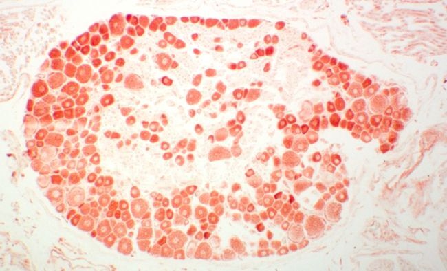
- Experimental details
- Immunohistochemistry of ET1 in mouse dorsal root ganglia