Explore
 Validate
 Learn
Learn Western blot
 Immunohistochemistry
Antibody data
- Antibody Data
- Antigen structure
- References [1]
- Comments [0]
- Validations
- Western blot [1]
- Immunocytochemistry [1]
- Flow cytometry [1]
Submit
Validation data
Reference
Comment
Report error
- Product number
- GTX22786 - Provider product page

- Provider
- GeneTex
- Proper citation
- GeneTex Cat#GTX22786, RRID:AB_384849
- Product name
- Endothelin 1 antibody [TR.ET.48.5]
- Antibody type
- Monoclonal
- Reactivity
- Human, Mouse, Rat, Canine, Porcine, Sheep
- Host
- Mouse
Submitted references Increased tissue endothelin-1 and endothelin-B receptor expression in temporal arteries from patients with giant cell arteritis.
Dimitrijevic I, Andersson C, Rissler P, Edvinsson L
Ophthalmology 2010 Mar;117(3):628-36
Ophthalmology 2010 Mar;117(3):628-36
No comments: Submit comment
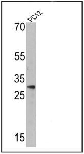
Supportive validation
- Submitted by
- GeneTex (provider)
- Main image

- Experimental details
- Western blot analysis of Endothelin 1 in 25 ug of PC12 cell lysates. Proteins were transferred to a PVDF membrane and blocked at 4¢XC overnight. The membrane was probed with Endothelin 1 antibody [TR.ET.48.5] at a dilution of 1:500 overnight at 4¢XC, washed in TBST, and probed with an HRP-conjugated secondary antibody for 1 hr at room temperature in the dark. Chemiluminescent detection was performed.
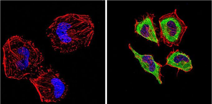
Supportive validation
- Submitted by
- GeneTex (provider)
- Main image

- Experimental details
- Immunofluorescent analysis of Endothelin 1 (green) in HeLa cells compared with a negative control in the absence of primary antibody (left). Formalin-fixed cells were permeabilized with 0.1% Triton X-100 in TBS for 5-10 minutes, blocked with 3% BSA-PBS for 30 minutes at room temperature and probed with Endothelin 1 antibody [TR.ET.48.5] in 3% BSA-PBS at a dilution of 1:200 and incubated overnight at 4¢XC in a humidified chamber. Cells were washed with PBST and incubated with a proper secondary antibody in PBS at room temperature in the dark. F-actin (red) was stained with a flourescent red phalloidin and nuclei (blue) were stained with DAPI for 5-10 minutes in the dark. Images were taken at a magnification of 60x.
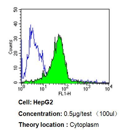
Supportive validation
- Submitted by
- GeneTex (provider)
- Main image
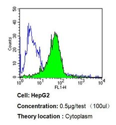
- Experimental details
- Flow cytometry analysis of Endothelin 1 in HepG2 cells compared to an isotype control (blue). Cells were harvested, adjusted to a concentration of 1-5x10^6 cells/ml, fixed with 2% paraformaldehyde, washed with PBS, and incubated with Endothelin 1 antibody [TR.ET.48.5] at a dilution of 0.5 ug/test for 60 min at room temperature. Cells were then blocked in a solution of 2% BSA-PBS for 30 min at room temperature, incubated for 40 min at room temperature in the dark using a proper secondary antibody, and re-suspended in PBS for FACS analysis.