 Explore
 Validate
 Learn
Learn Western blot
 Immunocytochemistry
Antibody data
- Antibody Data
- Antigen structure
- References [2]
- Comments [0]
- Validations
- Immunocytochemistry [8]
Submit
Validation data
Reference
Comment
Report error
- Product number
- MA3-941 - Provider product page

- Provider
- Invitrogen Antibodies
- Product name
- Mu-Calpain Monoclonal Antibody (2H2A7C2)
- Antibody type
- Monoclonal
- Antigen
- Purifed from natural sources
- Description
- MA3-941 detects mu-calpain from human platelets and erythrocytes, bovine platelets, heart and skeletal muscle, rat myoblasts, kidney, liver and spleen, and pig cultured cells. This antibody does not cross-react with m-calpain, n-calpain, calmodulin or calpastatin. MA3-941 has been successfully used in Western blot, IF and immunocytochemistry procedures. By Western blot, this antibody detects an ~80 kDa protein representing mu-calpain from human platelets and erythrocytes. Immunocytochemical staining of mu-calpain in porcine LLC-PK1 cells with MA3-941 results in diffuse cytoplasmic staining. This product has not been shown to be effective in immunoprecipitation experiments. The MA3-941 antigen is purified bovine skeletal muscle 80 kDa mu-calpain subunit. This antibody recognizes an epitope between amino acids 245-265 (domain II) of human mu-calpain.
- Reactivity
- Human, Rat, Bovine, Porcine
- Host
- Mouse
- Isotype
- IgG
- Antibody clone number
- 2H2A7C2
- Vial size
- 100 μL
- Concentration
- Conc. Not Determined
- Storage
- -20°C, Avoid Freeze/Thaw Cycles
Submitted references Effect of monoclonal antibodies specific for the 28-kDa subunit on catalytic properties of the calpains.
A comparison of the intracellular distribution of mu-calpain, m-calpain, and calpastatin in proliferating human A431 cells.
Cong J, Thompson VF, Goll DE
The Journal of biological chemistry 1993 Dec 5;268(34):25740-7
The Journal of biological chemistry 1993 Dec 5;268(34):25740-7
A comparison of the intracellular distribution of mu-calpain, m-calpain, and calpastatin in proliferating human A431 cells.
Lane RD, Allan DM, Mellgren RL
Experimental cell research 1992 Nov;203(1):5-16
Experimental cell research 1992 Nov;203(1):5-16
No comments: Submit comment
Supportive validation
- Submitted by
- Invitrogen Antibodies (provider)
- Main image
- Experimental details
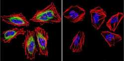
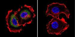
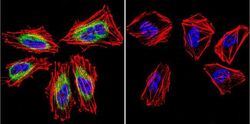
- Immunofluorescent analysis of Mu-Calpain in HeLa Cells. Cells were grown on chamber slides and fixed with formaldehyde prior to staining. Cells were probed without (control) or with a Mu-Calpain monoclonal antibody (Product # MA3-941) at a dilution of 1:20 overnight at 4 C, washed with PBS and incubated with a DyLight-488 conjugated secondary antibody (Product # 35503). Mu-Calpain staining (green), F-Actin staining with Phalloidin (red) and nuclei with DAPI (blue) is shown. Images were taken at 60X magnification.

- Submitted by
- Invitrogen Antibodies (provider)
- Main image
- Experimental details
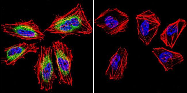
- Immunofluorescent analysis of Mu-Calpain in MCF-7 Cells. Cells were grown on chamber slides and fixed with formaldehyde prior to staining. Cells were probed without (control) or with a Mu-Calpain monoclonal antibody (Product # MA3-941) at a dilution of 1:20 overnight at 4 C, washed with PBS and incubated with a DyLight-488 conjugated secondary antibody (Product # 35503). Mu-Calpain staining (green), F-Actin staining with Phalloidin (red) and nuclei with DAPI (blue) is shown. Images were taken at 60X magnification.
- Submitted by
- Invitrogen Antibodies (provider)
- Main image

- Experimental details
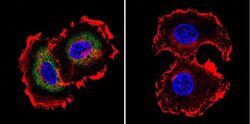
- Immunofluorescent analysis of Mu-Calpain in U251 Cells. Cells were grown on chamber slides and fixed with formaldehyde prior to staining. Cells were probed without (control) or with a Mu-Calpain monoclonal antibody (Product # MA3-941) at a dilution of 1:20 overnight at 4 C, washed with PBS and incubated with a DyLight-488 conjugated secondary antibody (Product # 35503). Mu-Calpain staining (green), F-Actin staining with Phalloidin (red) and nuclei with DAPI (blue) is shown. Images were taken at 60X magnification.
- Submitted by
- Invitrogen Antibodies (provider)
- Main image

- Experimental details
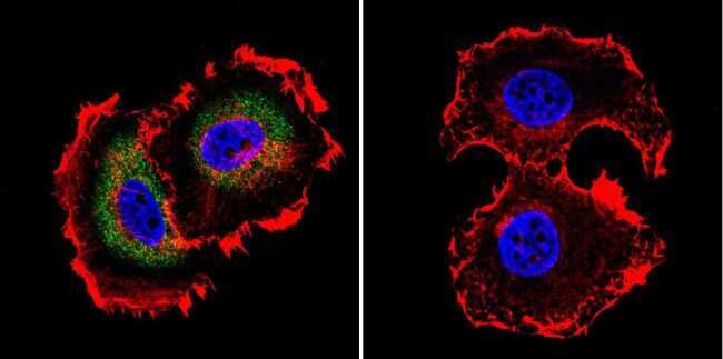
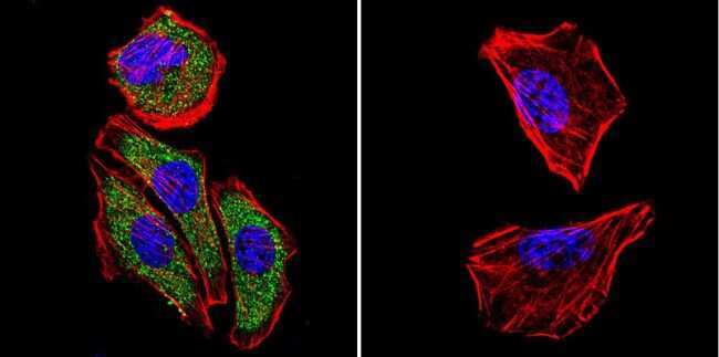
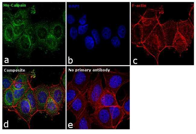
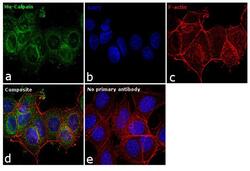
- Immunofluorescence analysis of Mu-Calpain was performed using 70% confluent log phase Hep G2 cells. The cells were fixed with 4% paraformaldehyde for 10 minutes, permeabilized with 0.1% Triton™ X-100 for 15 minutes, and blocked with 1% BSA for 1 hour at room temperature. The cells were labeled with Mu-Calpain (2H2A7C2) Monoclonal Antibody (Product # MA3-941) at 1:20 dilution in 0.1% BSA, incubated at 4 degree Celsius overnight and then labeled with Goat anti-Mouse IgG (H+L) Superclonal™ Secondary Antibody, Alexa Fluor® 488 conjugate (Product # A28175) at a dilution of 1:2000 for 45 minutes at room temperature (Panel a: green). Nuclei (Panel b: blue) were stained with SlowFade® Gold Antifade Mountant with DAPI (Product # S36938). F-actin (Panel c: red) was stained with Rhodamine Phalloidin (Product # R415, 1:300). Panel d represents the merged image showing cytoplasmic localization. Panel e represents control cells with no primary antibody to assess background. The images were captured at 60X magnification.
- Submitted by
- Invitrogen Antibodies (provider)
- Main image
- Experimental details
- Immunofluorescent analysis of Mu-Calpain in HeLa Cells. Cells were grown on chamber slides and fixed with formaldehyde prior to staining. Cells were probed without (control) or with a Mu-Calpain monoclonal antibody (Product # MA3-941) at a dilution of 1:20 overnight at 4 C, washed with PBS and incubated with a DyLight-488 conjugated secondary antibody (Product # 35503). Mu-Calpain staining (green), F-Actin staining with Phalloidin (red) and nuclei with DAPI (blue) is shown. Images were taken at 60X magnification.
- Submitted by
- Invitrogen Antibodies (provider)
- Main image
- Experimental details
- Immunofluorescent analysis of Mu-Calpain in MCF-7 Cells. Cells were grown on chamber slides and fixed with formaldehyde prior to staining. Cells were probed without (control) or with a Mu-Calpain monoclonal antibody (Product # MA3-941) at a dilution of 1:20 overnight at 4 C, washed with PBS and incubated with a DyLight-488 conjugated secondary antibody (Product # 35503). Mu-Calpain staining (green), F-Actin staining with Phalloidin (red) and nuclei with DAPI (blue) is shown. Images were taken at 60X magnification.

- Submitted by
- Invitrogen Antibodies (provider)
- Main image

- Experimental details
- Immunofluorescent analysis of Mu-Calpain in U251 Cells. Cells were grown on chamber slides and fixed with formaldehyde prior to staining. Cells were probed without (control) or with a Mu-Calpain monoclonal antibody (Product # MA3-941) at a dilution of 1:20 overnight at 4 C, washed with PBS and incubated with a DyLight-488 conjugated secondary antibody (Product # 35503). Mu-Calpain staining (green), F-Actin staining with Phalloidin (red) and nuclei with DAPI (blue) is shown. Images were taken at 60X magnification.

- Submitted by
- Invitrogen Antibodies (provider)
- Main image
- Experimental details
- Immunofluorescence analysis of Mu-Calpain was performed using 70% confluent log phase Hep G2 cells. The cells were fixed with 4% paraformaldehyde for 10 minutes, permeabilized with 0.1% Triton™ X-100 for 15 minutes, and blocked with 1% BSA for 1 hour at room temperature. The cells were labeled with Mu-Calpain (2H2A7C2) Monoclonal Antibody (Product # MA3-941) at 1:20 dilution in 0.1% BSA, incubated at 4 degree Celsius overnight and then labeled with Goat anti-Mouse IgG (H+L) Superclonal™ Secondary Antibody, Alexa Fluor® 488 conjugate (Product # A28175) at a dilution of 1:2000 for 45 minutes at room temperature (Panel a: green). Nuclei (Panel b: blue) were stained with SlowFade® Gold Antifade Mountant with DAPI (Product # S36938). F-actin (Panel c: red) was stained with Rhodamine Phalloidin (Product # R415, 1:300). Panel d represents the merged image showing cytoplasmic localization. Panel e represents control cells with no primary antibody to assess background. The images were captured at 60X magnification.
