Explore
 Validate
 Learn
LearnMAB817
antibody from Novus Biologicals
Targeting: BIRC3
API2, c-IAP2, cIAP2, hiap-1, MALT2, MIHC, RNF49
 Western blot
Antibody data
- Antibody Data
- Antigen structure
- References [5]
- Comments [0]
- Validations
- Western blot [2]
- Immunohistochemistry [2]
Submit
Validation data
Reference
Comment
Report error
- Product number
- MAB817 - Provider product page

- Provider
- Novus Biologicals
- Product name
- Mouse Monoclonal cIAP-2/HIAP-1 Antibody
- Antibody type
- Monoclonal
- Description
- Protein A or G purified from hybridoma culture supernatant. Detects human and mouse cIAP-2/HIAP-1 in Western blots. Does not cross-react with HEK293 cells transfected with cIAP-1.
- Reactivity
- Human, Mouse
- Host
- Mouse
- Isotype
- IgG
- Vial size
- 100 ug
- Concentration
- LYOPH
- Storage
- Use a manual defrost freezer and avoid repeated freeze-thaw cycles. 12 months from date of receipt, -20 to -70 degreesC as supplied. 1 month, 2 to 8 degreesC under sterile conditions after reconstitution. 6 months, -20 to -70 degreesC under sterile conditions after reconstitution.
Submitted references Close Relationship between cIAP2 and Human ARDS Induced by Severe H7N9 Infection.
Dendritic Cell RIPK1 Maintains Immune Homeostasis by Preventing Inflammation and Autoimmunity.
BIRC3 is a biomarker of mesenchymal habitat of glioblastoma, and a mediator of survival adaptation in hypoxia-driven glioblastoma habitats.
Enhanced susceptibility to tumor necrosis factor-related apoptosis-inducing ligand-mediated apoptosis in oral squamous cell carcinoma cells treated with phosphatidylinositol 3-kinase inhibitors.
Effect of hyperthermia on TRAIL-induced apoptotic death in human colon cancer cells: development of a novel strategy for regional therapy.
Qin C, Sai XY, Qian XF, Wu Y, Zou LF, Wang HM, Bian T, Yan Z
BioMed research international 2019;2019:2121357
BioMed research international 2019;2019:2121357
Dendritic Cell RIPK1 Maintains Immune Homeostasis by Preventing Inflammation and Autoimmunity.
O'Donnell JA, Lehman J, Roderick JE, Martinez-Marin D, Zelic M, Doran C, Hermance N, Lyle S, Pasparakis M, Fitzgerald KA, Marshak-Rothstein A, Kelliher MA
Journal of immunology (Baltimore, Md. : 1950) 2018 Jan 15;200(2):737-748
Journal of immunology (Baltimore, Md. : 1950) 2018 Jan 15;200(2):737-748
BIRC3 is a biomarker of mesenchymal habitat of glioblastoma, and a mediator of survival adaptation in hypoxia-driven glioblastoma habitats.
Wang D, Berglund AE, Kenchappa RS, MacAulay RJ, Mulé JJ, Etame AB
Scientific reports 2017 Aug 24;7(1):9350
Scientific reports 2017 Aug 24;7(1):9350
Enhanced susceptibility to tumor necrosis factor-related apoptosis-inducing ligand-mediated apoptosis in oral squamous cell carcinoma cells treated with phosphatidylinositol 3-kinase inhibitors.
Uchida M, Iwase M, Takaoka S, Yoshiba S, Kondo G, Watanabe H, Ohashi M, Nagumo M, Shintani S
International journal of oncology 2007 May;30(5):1163-71
International journal of oncology 2007 May;30(5):1163-71
Effect of hyperthermia on TRAIL-induced apoptotic death in human colon cancer cells: development of a novel strategy for regional therapy.
Yoo J, Lee YJ
Journal of cellular biochemistry 2007 Jun 1;101(3):619-30
Journal of cellular biochemistry 2007 Jun 1;101(3):619-30
No comments: Submit comment
Supportive validation
- Submitted by
- Novus Biologicals (provider)
- Main image

- Experimental details
- Detection of Human and Mouse cIAP-2/HIAP-1 by Western Blot. Western blot shows lysates of Raji human Burkitt's lymphoma cell line, Daudi human Burkitt's lymphoma cell line, and A20 mouse B cell lymphoma cell line. PVDF membrane was probed with 0.5 µg/mL of Mouse Anti-Human/Mouse cIAP-2/HIAP-1 Monoclonal Antibody (Catalog # MAB817) followed by HRP-conjugated Anti-Mouse IgG Secondary Antibody (Catalog # HAF007). A specific band was detected for cIAP-2/HIAP-1 at approximately 68 kDa (as indicated). This experiment was conducted under reducing conditions and using Immunoblot Buffer Group 5.

- Submitted by
- Novus Biologicals (provider)
- Main image

- Experimental details
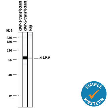
- Detection of Human cIAP-2/HIAP-1 by Simple WesternTM. Simple Western lane view shows lysates of HEK293 human embryonic kidney cell line transfected with either cIAP-1 or cIAP-2 and Raji human Burkitt's lymphoma cell line, loaded at 0.2 mg/mL. A specific band was detected for cIAP-2/HIAP-1 at approximately 74 kDa (as indicated) using 10 µg/mL of Mouse Anti-Human/Mouse cIAP-2/HIAP-1 Monoclonal Antibody (Catalog # MAB817). This experiment was conducted under reducing conditions and using the 12-230 kDa separation system.
Supportive validation
- Submitted by
- Novus Biologicals (provider)
- Main image
- Experimental details
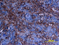
- cIAP-2/HIAP-1 in Human Colon Cancer Tissue. cIAP-2/HIAP-1 was detected in immersion fixed paraffin-embedded sections of human colon cancer tissue using Mouse Anti-Human/Mouse cIAP-2/ HIAP-1 Monoclonal Antibody (Catalog # MAB817) at 25 µg/mL overnight at 4 °C. Tissue was stained using the Anti-Mouse HRP-DAB Cell & Tissue Staining Kit (brown; Catalog # CTS002) and counter-stained with hematoxylin (blue). View our protocol for Chromogenic IHC Staining of Paraffin-embedded Tissue Sections. This application has not been tested in mouse samples.

- Submitted by
- Novus Biologicals (provider)
- Main image

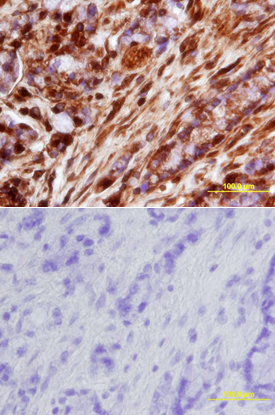
- Experimental details
- cIAP-2/HIAP-1 in Human Colon. cIAP-2/HIAP-1 was detected in immersion fixed paraffin-embedded sections of human colon array using Mouse Anti-Human/Mouse cIAP-2/HIAP-1 Monoclonal Antibody (Catalog # MAB817) at 25 µg/mL overnight at 4 °C. Tissue was stained using the Anti-Mouse HRP-DAB Cell & Tissue Staining Kit (brown; Catalog # CTS002) and counterstained with hematoxylin (blue). Lower panel shows a lack of labeling if primary antibodies are omitted and tissue is stained only with secondary antibody followed by incubation with detection reagents. View our protocol for Chromogenic IHC Staining of Paraffin-embedded Tissue Sections. This application has not been tested in mouse samples.