 Explore
 Validate
 Learn
LearnPA5-19514
antibody from Invitrogen Antibodies
Targeting: BIRC3
API2, c-IAP2, cIAP2, hiap-1, MALT2, MIHC, RNF49
 Western blot
 Immunocytochemistry
Antibody data
- Antibody Data
- Antigen structure
- References [0]
- Comments [0]
- Validations
- Immunocytochemistry [1]
Submit
Validation data
Reference
Comment
Report error
- Product number
- PA5-19514 - Provider product page

- Provider
- Invitrogen Antibodies
- Product name
- cIAP2 Polyclonal Antibody
- Antibody type
- Polyclonal
- Antigen
- Synthetic peptide
- Description
- For Western Blot, this antibody has non-specific bands at 150 kDa,35 kDa and 75 kDa. This antibody is predicted to react with mouse, rat, chicken and dog based on sequence homology.
- Reactivity
- Human, Rat
- Host
- Rabbit
- Isotype
- IgG
- Vial size
- 100 μg
- Concentration
- 0.5 mg/mL
- Storage
- Store at 4°C short term. For long term storage, store at -20°C, avoiding freeze/thaw cycles.
No comments: Submit comment
Supportive validation
- Submitted by
- Invitrogen Antibodies (provider)
- Main image
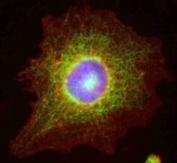
- Experimental details
- Immunofluorescent staining of HeLa cells using Product # PA5-19514, anti-cIAP2 antibody. The cells were fixed with methanol (100%) for 5 minutes and exposed to the primary antibody at a concentration of 1 µg/mL for 1 hour at room temp. The secondary antibody was a 448 fluorescence conjugated Goat anti-rabbit IgG (green) at a dilution of 1:1000. A WGA- 594 fluorescent conjugated stain was used to label plasma membranes (red) and the nuclei stain was DAPI (blue).
