 Explore
 Validate
 Learn
Learn Western blot
 ELISA
ELISA Immunoprecipitation
Antibody data
- Antibody Data
- Antigen structure
- References [0]
- Comments [0]
- Validations
- Western blot [2]
- Immunoprecipitation [2]
- Immunohistochemistry [1]
Submit
Validation data
Reference
Comment
Report error
- Product number
- LS-B10634 - Provider product page

- Provider
- LSBio
- Product name
- IHC-plus™ TPMT Antibody LS-B10634
- Antibody type
- Polyclonal
- Description
- Caprylic acid and ammonium sulfate precipitation
- Reactivity
- Human
- Host
- Rabbit
- Isotype
- IgG
- Storage
- Aliquot and store at -20°C or -80°C. Avoid freeze-thaw cycles.
No comments: Submit comment
Enhanced validation
- Submitted by
- LSBio (provider)
- Enhanced method
- Genetic validation
- Main image
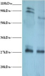
- Experimental details
- Western blot of Thiopurine S-methyltransferase antibody at 2 ug/ml. Lane 1: EC109 whole cell lysate. Lane 2: 293T whole cell lysate. Secondary: Goat polyclonal to Rabbit IgG at 1:15000 dilution. Predicted band size: 27 kDa. Observed band size: 27 kDa Additional bands at: 90 kDa, 100 kDa. We are unsure as to the identity of these extra bands. This image was taken for the unconjugated form of this product. Other forms have not been tested.

- Submitted by
- LSBio (provider)
- Enhanced method
- Genetic validation
- Main image

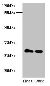
- Experimental details
- Western blot All lanes: Thiopurine S-methyltransferase antibody at 2µg/ml Lane 1: EC109 whole cell lysate Lane 2: 293T whole cell lysate Secondary Goat polyclonal to rabbit IgG at 1/15000 dilution Predicted band size: 27 kDa Observed band size: 27 kDa
Supportive validation
- Submitted by
- LSBio (provider)
- Enhanced method
- Genetic validation
- Main image
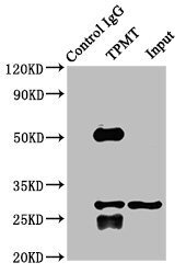
- Experimental details
- Immunoprecipitating TPMT in K562 whole cell lysate Lane 1: Rabbit control IgG instead of TPMT Antibody in K562 whole cell lysate.For western blotting, a HRP-conjugated Protein G antibody was used as the secondary antibody (1/2000) Lane 2: TPMT Antibody (8µg) + K562 whole cell lysate (500µg) Lane 3: K562 whole cell lysate (10µg)

- Submitted by
- LSBio (provider)
- Main image

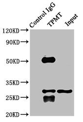
- Experimental details
- Immunoprecipitating TPMT in K562 whole cell lysate Lane 1: Rabbit control IgG instead of TPMT Antibody in K562 whole cell lysate.For western blotting, a HRP-conjugated Protein G antibody was used as the secondary antibody (1/2000) Lane 2: TPMT Antibody (8µg) + K562 whole cell lysate (500µg) Lane 3: K562 whole cell lysate (10µg)
Supportive validation
- Submitted by
- LSBio (provider)
- Enhanced method
- Genetic validation
- Main image
- Experimental details
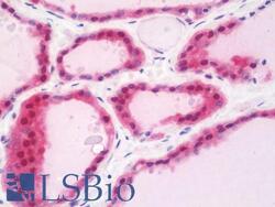
- Anti-TPMT antibody IHC staining of human thyroid. Immunohistochemistry of formalin-fixed, paraffin-embedded tissue after heat-induced antigen retrieval. Antibody concentration 10 ug/ml. This image was taken for the unconjugated form of this product. Other forms have not been tested.
