 Explore
 Validate
 Learn
Learn Western blot
Antibody data
- Antibody Data
- Antigen structure
- References [0]
- Comments [0]
- Validations
- Western blot [2]
- Immunocytochemistry [5]
- Immunoprecipitation [2]
- Other assay [1]
Submit
Validation data
Reference
Comment
Report error
- Product number
- PA5-117919 - Provider product page

- Provider
- Invitrogen Antibodies
- Product name
- CRMP2 Polyclonal Antibody
- Antibody type
- Polyclonal
- Antigen
- Other
- Reactivity
- Human
- Host
- Rabbit
- Isotype
- IgG
- Vial size
- 100 μL
- Storage
- -20°C, Avoid Freeze/Thaw Cycles
No comments: Submit comment
Supportive validation
- Submitted by
- Invitrogen Antibodies (provider)
- Main image
- Experimental details
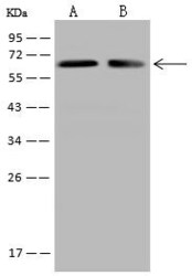
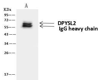
- Western Blot using CRMP2 Polyclonal Antibody (Product # PA5-117919) at 1:500 dilution. Lane A: Jurkat Whole Cell Lysate, Lane B: U87MG Whole Cell Lysate. Lysates/proteins at 30 μg per lane. Secondary antibody: Goat Anti-Rabbit IgG (H+L)/HRP at 1:10,000 dilution. Developed using the ECL technique. Performed under reducing conditions. Predicted band size: 62 kDa. Observed band size: 62 kDa.
- Submitted by
- Invitrogen Antibodies (provider)
- Main image
- Experimental details
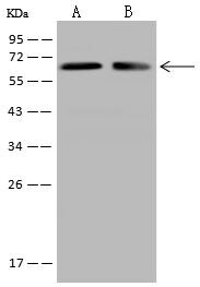
- Western Blot of CRMP2 in Lane A: Jurkat Whole Cell Lysate, Lane B: U87MG Whole Cell Lysate. Samples (30 µg per lane) were incubated with polyclonal antibody (Product # PA5-117919) with a dilution of 1:500 , followed by Goat Anti-Rabbit IgG (H+L)/HRP using a dilution of 1:10,000. Assay was performed under reducing conditions. Predicted band size: 62 kDa, Observed band size: 62 kDa.

Supportive validation
- Submitted by
- Invitrogen Antibodies (provider)
- Main image

- Experimental details
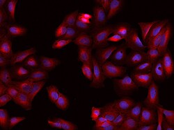
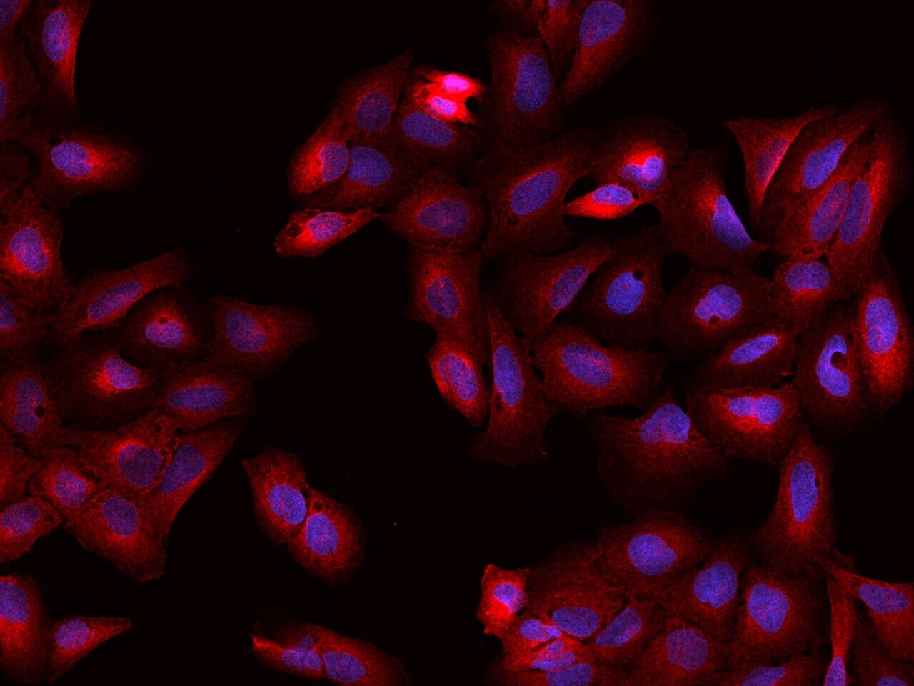
- Immunofluorescence staining of CRMP2 in U2OS cells. Cells were fixed with 4% PFA, permeabilzed with 0.1% Triton X-100 in PBS, blocked with 10% serum, and incubated with CRMP2 Polyclonal Antibody (Product # PA5-117919, 1:200) at 4°C overnight. Then cells were stained with the Alexa Fluor®594-conjugated Goat Anti-rabbit IgG secondary antibody (red) and counterstained with DAPI (blue). Positive staining was localized to cytoplasm.

- Submitted by
- Invitrogen Antibodies (provider)
- Main image

- Experimental details
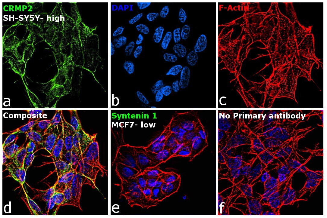
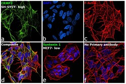
- Immunofluorescence analysis of DPYSL2 was performed using 70% confluent log phase SH-SY5Y and MCF7 cells. The cells were fixed with 4% paraformaldehyde for 10 minutes, permeabilized with 0.1% Triton™ X-100 for 15 minutes, and blocked with 2% BSA for 1 hour at room temperature. The cells were labeled with CRMP2 Polyclonal Antibody (Product # PA5-117919) at 1:100 dilution in 0.1% BSA, incubated at 4 degree celsius overnight and then labeled with Donkey anti-Rabbit IgG (H+L) Highly Cross-Adsorbed Secondary Antibody, Alexa Fluor Plus 488 (Product # A32790), (1:2000 dilution), for 45 minutes at room temperature (Panel a: Green). Nuclei (Panel b: Blue) were stained with ProLong™ Diamond Antifade Mountant with DAPI (Product # P36962). F-actin (Panel c: Red) was stained with Rhodamine Phalloidin (Product # R415, 1:300). Panel d represents the merged image showing cytoplasmic localization. Panel e represents MCF7 cells with low expression of CRMP2 protein. Panel f represents control cells with no primary antibody to assess background. The images were captured at 60X magnification.
- Submitted by
- Invitrogen Antibodies (provider)
- Main image
- Experimental details
- Immunocytochemistry analysis of CRMP2 in U2OS cells. Samples were treated with 4% PFA, permeabilzed with 0.1% Triton X-100 in PBS, blocked with 10% serum, incubated with polyclonal antibody (Product # PA5-117919) with a dilution of 1: 200 (4°C overnight), followed by Alexa Fluor 594-conjugated Goat Anti-rabbit IgG secondary antibody (red).

- Submitted by
- Invitrogen Antibodies (provider)
- Main image

- Experimental details
- Immunofluorescence staining of CRMP2 in U2OS cells. Cells were fixed with 4% PFA, permeabilzed with 0.1% Triton X-100 in PBS, blocked with 10% serum, and incubated with CRMP2 Polyclonal Antibody (Product # PA5-117919, 1:200) at 4°C overnight. Then cells were stained with the Alexa Fluor®594-conjugated Goat Anti-rabbit IgG secondary antibody (red) and counterstained with DAPI (blue). Positive staining was localized to cytoplasm.
- Submitted by
- Invitrogen Antibodies (provider)
- Main image
- Experimental details
- Immunofluorescence analysis of DPYSL2 was performed using 70% confluent log phase SH-SY5Y and MCF7 cells. The cells were fixed with 4% paraformaldehyde for 10 minutes, permeabilized with 0.1% Triton™ X-100 for 15 minutes, and blocked with 2% BSA for 1 hour at room temperature. The cells were labeled with CRMP2 Polyclonal Antibody (Product # PA5-117919) at 1:100 dilution in 0.1% BSA, incubated at 4 degree celsius overnight and then labeled with Donkey anti-Rabbit IgG (H+L) Highly Cross-Adsorbed Secondary Antibody, Alexa Fluor Plus 488 (Product # A32790), (1:2000 dilution), for 45 minutes at room temperature (Panel a: Green). Nuclei (Panel b: Blue) were stained with ProLong™ Diamond Antifade Mountant with DAPI (Product # P36962). F-actin (Panel c: Red) was stained with Rhodamine Phalloidin (Product # R415, 1:300). Panel d represents the merged image showing cytoplasmic localization. Panel e represents MCF7 cells with low expression of CRMP2 protein. Panel f represents control cells with no primary antibody to assess background. The images were captured at 60X magnification.

Supportive validation
- Submitted by
- Invitrogen Antibodies (provider)
- Main image

- Experimental details
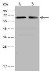
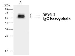
- CRMP2 Immunoprecipitation using: Lane A: 0.5 mg Jurkat Whole Cell Lysate 4 µL with CRMP2 Polyclonal Antibody (Product # PA5-117919) and 60 μg of Immunomagnetic beads Protein A/G. Primary antibody: CRMP2 Polyclonal Antibody, at 1:100 dilution. Secondary antibody: Goat Anti-Rabbit IgG (H+L) /HRP at 1:10,000 dilution. Developed using the ECL technique. Performed under reducing conditions. Predicted band size: 62 kDa. Observed band size: 62 kDa.

- Submitted by
- Invitrogen Antibodies (provider)
- Main image

- Experimental details
- Immunoprecipitation of CRMP2 in Lane A: 0.5 mg Jurkat Whole Cell Lysate. Samples were treated with 60 μg of immunomagnetic Protein A/G beads, incubated with polyclonal antibody (Product # PA5-117919) with a dilution of 1:100 , followed by Goat Anti-Rabbit IgG (H+L)/HRP using a dilution of 1:10,000. Assay was performed under reducing conditions. Predicted band size: 62 kDa , Observed band size : 62 kDa .
Supportive validation
- Submitted by
- Invitrogen Antibodies (provider)
- Main image
- Experimental details
- CRMP2 Immunoprecipitation using: Lane A: 0.5 mg Jurkat Whole Cell Lysate 4 µL with CRMP2 Polyclonal Antibody (Product # PA5-117919) and 60 μg of Immunomagnetic beads Protein A/G. Primary antibody: CRMP2 Polyclonal Antibody, at 1:100 dilution. Secondary antibody: Goat Anti-Rabbit IgG (H+L) /HRP at 1:10,000 dilution. Developed using the ECL technique. Performed under reducing conditions. Predicted band size: 62 kDa. Observed band size: 62 kDa.
