 Explore
 Validate
 Learn
Learn Western blot
 Immunohistochemistry
Antibody data
- Antibody Data
- Antigen structure
- References [4]
- Comments [0]
- Validations
- Immunohistochemistry [1]
Submit
Validation data
Reference
Comment
Report error
- Product number
- AF1013 - Provider product page

- Provider
- R&D Systems
- Product name
- Mouse Cathepsin H Antibody
- Antibody type
- Polyclonal
- Description
- Antigen Affinity-purified. Detects mouse Cathepsin H in direct ELISAs and Western blots. In direct ELISAs, less than 2% cross-reactivity with recombinant mouse (rm) Cathepsin L, and rmCathepsin J is observed.
- Reactivity
- Mouse
- Host
- Goat
- Conjugate
- Unconjugated
- Antigen sequence
Q3UCD6- Isotype
- IgG
- Vial size
- 100 ug
- Concentration
- LYOPH
- Storage
- Use a manual defrost freezer and avoid repeated freeze-thaw cycles. 12 months from date of receipt, -20 to -70 °C as supplied. 1 month, 2 to 8 °C under sterile conditions after reconstitution. 6 months, -20 to -70 °C under sterile conditions after reconstitution.
Submitted references Lysosomal protein turnover contributes to the acquisition of TGFβ-1 induced invasive properties of mammary cancer cells.
Alveolar progenitor and stem cells in lung development, renewal and cancer.
Gene targeting of the cysteine peptidase cathepsin H impairs lung surfactant in mice.
Macrophages and cathepsin proteases blunt chemotherapeutic response in breast cancer.
Kern U, Wischnewski V, Biniossek ML, Schilling O, Reinheckel T
Molecular cancer 2015 Feb 15;14:39
Molecular cancer 2015 Feb 15;14:39
Alveolar progenitor and stem cells in lung development, renewal and cancer.
Desai TJ, Brownfield DG, Krasnow MA
Nature 2014 Mar 13;507(7491):190-4
Nature 2014 Mar 13;507(7491):190-4
Gene targeting of the cysteine peptidase cathepsin H impairs lung surfactant in mice.
Bühling F, Kouadio M, Chwieralski CE, Kern U, Hohlfeld JM, Klemm N, Friedrichs N, Roth W, Deussing JM, Peters C, Reinheckel T
PloS one 2011;6(10):e26247
PloS one 2011;6(10):e26247
Macrophages and cathepsin proteases blunt chemotherapeutic response in breast cancer.
Shree T, Olson OC, Elie BT, Kester JC, Garfall AL, Simpson K, Bell-McGuinn KM, Zabor EC, Brogi E, Joyce JA
Genes & development 2011 Dec 1;25(23):2465-79
Genes & development 2011 Dec 1;25(23):2465-79
No comments: Submit comment
Supportive validation
- Submitted by
- R&D Systems (provider)
- Main image
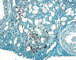
- Experimental details
- Cathepsin H in Mouse Ovary. Cathepsin H was detected in perfusion fixed frozen sections of mouse ovary using 1.7 µg/mL Goat Anti-Mouse Cathepsin H Antigen Affinity-purified Polyclonal Antibody (Catalog # AF1013) overnight at 4 °C. Tissue was stained with the Anti-Goat HRP-DAB Cell & Tissue Staining Kit (brown; Catalog # CTS008) and counterstained with hematoxylin (blue). View our protocol for Chromogenic IHC Staining of Frozen Tissue Sections.
