 Explore
 Validate
 Learn
Learn Western blot
 Immunocytochemistry
Antibody data
- Antibody Data
- Antigen structure
- References [20]
- Comments [0]
- Validations
- Western blot [1]
- Immunohistochemistry [3]
- Flow cytometry [1]
- Other assay [1]
Submit
Validation data
Reference
Comment
Report error
- Product number
- MA5-12731 - Provider product page

- Provider
- Invitrogen Antibodies
- Product name
- Anti-Cyclin D2 Antibody Cocktail
- Antibody type
- Monoclonal
- Antigen
- Recombinant full-length protein
- Description
- MA5-12731 targets Cyclin D2 in FACS, IF, and WB applications and shows reactivity with Human, mouse, and Rat samples. The MA5-12731 immunogen is purified histidine-tagged human recombinant full length cyclin D2 protein.
- Reactivity
- Human, Mouse, Rat
- Host
- Mouse
- Isotype
- IgG
- Antibody clone number
- DCS-3.1 + DCS-5.2
- Vial size
- 500 µL
- Concentration
- 0.2 mg/mL
- Storage
- 4° C
Submitted references XPO1 inhibitor combination therapy with bortezomib or carfilzomib induces nuclear localization of IκBα and overcomes acquired proteasome inhibitor resistance in human multiple myeloma.
De novo CCND2 mutations leading to stabilization of cyclin D2 cause megalencephaly-polymicrogyria-polydactyly-hydrocephalus syndrome.
Smoothened controls cyclin D2 expression and regulates the generation of intermediate progenitors in the developing cortex.
Sustained expression of the transcription factor GLIS3 is required for normal beta cell function in adults.
GSK-3 inactivation or depletion promotes β-cell replication via down regulation of the CDK inhibitor, p27 (Kip1).
GSK-3 inactivation or depletion promotes β-cell replication via down regulation of the CDK inhibitor, p27 (Kip1).
Cyclin D3 compensates for the loss of cyclin D1 during ErbB2-induced mammary tumor initiation and progression.
Cyclin D2 protein stability is regulated in pancreatic beta-cells.
Adaptive beta-cell proliferation is severely restricted with advanced age.
Selective cortical interneuron and GABA deficits in cyclin D2-null mice.
Loss of cyclin D1 impairs cerebellar development and suppresses medulloblastoma formation.
Cdk4 is indispensable for postnatal proliferation of the anterior pituitary.
Signal transducer and activator of transcription 5 activation is sufficient to drive transcriptional induction of cyclin D2 gene and proliferation of rat pancreatic beta-cells.
Cyclin D2 compensates for the loss of cyclin D1 in estrogen-induced mouse uterine epithelial cell proliferation.
v-Jun overrides the mitogen dependence of S-phase entry by deregulating retinoblastoma protein phosphorylation and E2F-pocket protein interactions as a consequence of enhanced cyclin E-cdk2 catalytic activity.
v-Jun overrides the mitogen dependence of S-phase entry by deregulating retinoblastoma protein phosphorylation and E2F-pocket protein interactions as a consequence of enhanced cyclin E-cdk2 catalytic activity.
Targeted disruption of CDK4 delays cell cycle entry with enhanced p27(Kip1) activity.
Targeted disruption of CDK4 delays cell cycle entry with enhanced p27(Kip1) activity.
Regulation of exit from quiescence by p27 and cyclin D1-CDK4.
Regulation of exit from quiescence by p27 and cyclin D1-CDK4.
Turner JG, Kashyap T, Dawson JL, Gomez J, Bauer AA, Grant S, Dai Y, Shain KH, Meads M, Landesman Y, Sullivan DM
Oncotarget 2016 Nov 29;7(48):78896-78909
Oncotarget 2016 Nov 29;7(48):78896-78909
De novo CCND2 mutations leading to stabilization of cyclin D2 cause megalencephaly-polymicrogyria-polydactyly-hydrocephalus syndrome.
Mirzaa G, Parry DA, Fry AE, Giamanco KA, Schwartzentruber J, Vanstone M, Logan CV, Roberts N, Johnson CA, Singh S, Kholmanskikh SS, Adams C, Hodge RD, Hevner RF, Bonthron DT, Braun KPJ, Faivre L, Rivière JB, St-Onge J, Gripp KW, Mancini GM, Pang K, Sweeney E, van Esch H, Verbeek N, Wieczorek D, Steinraths M, Majewski J, FORGE Canada Consortium., Boycot KM, Pilz DT, Ross ME, Dobyns WB, Sheridan EG
Nature genetics 2014 May;46(5):510-515
Nature genetics 2014 May;46(5):510-515
Smoothened controls cyclin D2 expression and regulates the generation of intermediate progenitors in the developing cortex.
Komada M, Iguchi T, Takeda T, Ishibashi M, Sato M
Neuroscience letters 2013 Jun 28;547:87-91
Neuroscience letters 2013 Jun 28;547:87-91
Sustained expression of the transcription factor GLIS3 is required for normal beta cell function in adults.
Yang Y, Chang BH, Chan L
EMBO molecular medicine 2013 Jan;5(1):92-104
EMBO molecular medicine 2013 Jan;5(1):92-104
GSK-3 inactivation or depletion promotes β-cell replication via down regulation of the CDK inhibitor, p27 (Kip1).
Stein J, Milewski WM, Hara M, Steiner DF, Dey A
Islets 2011 Jan-Feb;3(1):21-34
Islets 2011 Jan-Feb;3(1):21-34
GSK-3 inactivation or depletion promotes β-cell replication via down regulation of the CDK inhibitor, p27 (Kip1).
Stein J, Milewski WM, Hara M, Steiner DF, Dey A
Islets 2011 Jan-Feb;3(1):21-34
Islets 2011 Jan-Feb;3(1):21-34
Cyclin D3 compensates for the loss of cyclin D1 during ErbB2-induced mammary tumor initiation and progression.
Zhang Q, Sakamoto K, Liu C, Triplett AA, Lin WC, Rui H, Wagner KU
Cancer research 2011 Dec 15;71(24):7513-24
Cancer research 2011 Dec 15;71(24):7513-24
Cyclin D2 protein stability is regulated in pancreatic beta-cells.
He LM, Sartori DJ, Teta M, Opare-Addo LM, Rankin MM, Long SY, Diehl JA, Kushner JA
Molecular endocrinology (Baltimore, Md.) 2009 Nov;23(11):1865-75
Molecular endocrinology (Baltimore, Md.) 2009 Nov;23(11):1865-75
Adaptive beta-cell proliferation is severely restricted with advanced age.
Rankin MM, Kushner JA
Diabetes 2009 Jun;58(6):1365-72
Diabetes 2009 Jun;58(6):1365-72
Selective cortical interneuron and GABA deficits in cyclin D2-null mice.
Glickstein SB, Moore H, Slowinska B, Racchumi J, Suh M, Chuhma N, Ross ME
Development (Cambridge, England) 2007 Nov;134(22):4083-93
Development (Cambridge, England) 2007 Nov;134(22):4083-93
Loss of cyclin D1 impairs cerebellar development and suppresses medulloblastoma formation.
Pogoriler J, Millen K, Utset M, Du W
Development (Cambridge, England) 2006 Oct;133(19):3929-37
Development (Cambridge, England) 2006 Oct;133(19):3929-37
Cdk4 is indispensable for postnatal proliferation of the anterior pituitary.
Jirawatnotai S, Aziyu A, Osmundson EC, Moons DS, Zou X, Kineman RD, Kiyokawa H
The Journal of biological chemistry 2004 Dec 3;279(49):51100-6
The Journal of biological chemistry 2004 Dec 3;279(49):51100-6
Signal transducer and activator of transcription 5 activation is sufficient to drive transcriptional induction of cyclin D2 gene and proliferation of rat pancreatic beta-cells.
Friedrichsen BN, Richter HE, Hansen JA, Rhodes CJ, Nielsen JH, Billestrup N, Møldrup A
Molecular endocrinology (Baltimore, Md.) 2003 May;17(5):945-58
Molecular endocrinology (Baltimore, Md.) 2003 May;17(5):945-58
Cyclin D2 compensates for the loss of cyclin D1 in estrogen-induced mouse uterine epithelial cell proliferation.
Chen B, Pollard JW
Molecular endocrinology (Baltimore, Md.) 2003 Jul;17(7):1368-81
Molecular endocrinology (Baltimore, Md.) 2003 Jul;17(7):1368-81
v-Jun overrides the mitogen dependence of S-phase entry by deregulating retinoblastoma protein phosphorylation and E2F-pocket protein interactions as a consequence of enhanced cyclin E-cdk2 catalytic activity.
Clark W, Black EJ, MacLaren A, Kruse U, LaThangue N, Vogt PK, Gillespie DA
Molecular and cellular biology 2000 Apr;20(7):2529-42
Molecular and cellular biology 2000 Apr;20(7):2529-42
Targeted disruption of CDK4 delays cell cycle entry with enhanced p27(Kip1) activity.
Tsutsui T, Hesabi B, Moons DS, Pandolfi PP, Hansel KS, Koff A, Kiyokawa H
Molecular and cellular biology 1999 Oct;19(10):7011-9
Molecular and cellular biology 1999 Oct;19(10):7011-9
Targeted disruption of CDK4 delays cell cycle entry with enhanced p27(Kip1) activity.
Tsutsui T, Hesabi B, Moons DS, Pandolfi PP, Hansel KS, Koff A, Kiyokawa H
Molecular and cellular biology 1999 Oct;19(10):7011-9
Molecular and cellular biology 1999 Oct;19(10):7011-9
Regulation of exit from quiescence by p27 and cyclin D1-CDK4.
Ladha MH, Lee KY, Upton TM, Reed MF, Ewen ME
Molecular and cellular biology 1998 Nov;18(11):6605-15
Molecular and cellular biology 1998 Nov;18(11):6605-15
Regulation of exit from quiescence by p27 and cyclin D1-CDK4.
Ladha MH, Lee KY, Upton TM, Reed MF, Ewen ME
Molecular and cellular biology 1998 Nov;18(11):6605-15
Molecular and cellular biology 1998 Nov;18(11):6605-15
No comments: Submit comment
Supportive validation
- Submitted by
- Invitrogen Antibodies (provider)
- Main image
- Experimental details
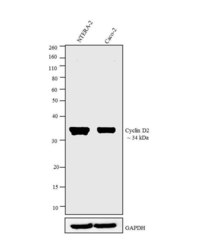
- Western blot analysis was performed on whole cell extracts (30 µg lysate) of NTERA-2 (Lane 1), and Caco-2 (Lane 2). The blots were probed with Anti-Cyclin D2 Mouse Monoclonal Antibody (Product # MA5-12731, 1-2 µg/mL) and detected by chemiluminescence using Goat anti-Mouse IgG (H+L) Secondary Antibody, HRP conjugate (Product # 62-6520, 1:4000 dilution). A 34 kDa band corresponding to Cyclin D2 was observed across cell lines tested. Known quantity of protein samples were electrophoresed using Novex® NuPAGE® 12 % Bis-Tris gel (Product # NP0342BOX), XCell SureLock™ Electrophoresis System (Product # EI0002) and Novex® Sharp Pre-Stained Protein Standard (Product # LC5800). Resolved proteins were then transferred onto a nitrocellulose membrane with iBlot® 2 Dry Blotting System (Product # IB21001). The membrane was probed with the relevant primary and secondary Antibody following blocking with 5 % skimmed milk. Chemiluminescent detection was performed using Pierce™ ECL Western Blotting Substrate (Product # 32106).

Supportive validation
- Submitted by
- Invitrogen Antibodies (provider)
- Main image
- Experimental details
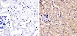
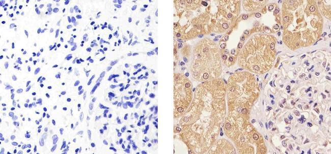
- Immunohistochemistry analysis of Cyclin D2 showing staining in the cytoplasm and nucleus of paraffin-embedded mouse kidney tissue (right) compared to a negative control without primary antibody (left). To expose target proteins, antigen retrieval was performed using 10mM sodium citrate (pH 6.0), microwaved for 8-15 min. Following antigen retrieval, tissues were blocked in 3% H2O2-methanol for 15 min at room temperature, washed with ddH2O and PBS, and then probed with a Cyclin D2 Antibody Mouse Monoclonal Antibody (Product # MA5-12731) diluted in 3% BSA-PBS at a dilution of 1:20 for 1 hour at 37°C in a humidified chamber. Tissues were washed extensively in PBST and detection was performed using an HRP-conjugated secondary antibody followed by colorimetric detection using a DAB kit. Tissues were counterstained with hematoxylin and dehydrated with ethanol and xylene to prep for mounting.
- Submitted by
- Invitrogen Antibodies (provider)
- Main image
- Experimental details
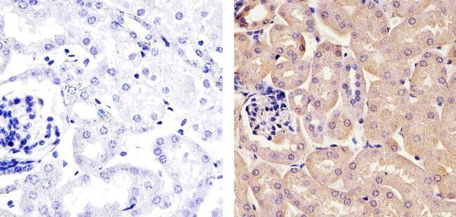
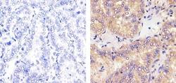
- Immunohistochemistry analysis of Cyclin D2 showing staining in the cytoplasm and nucleus of paraffin-embedded human kidney tissue (right) compared to a negative control without primary antibody (left). To expose target proteins, antigen retrieval was performed using 10mM sodium citrate (pH 6.0), microwaved for 8-15 min. Following antigen retrieval, tissues were blocked in 3% H2O2-methanol for 15 min at room temperature, washed with ddH2O and PBS, and then probed with a Cyclin D2 Antibody Mouse Monoclonal Antibody (Product # MA5-12731) diluted in 3% BSA-PBS at a dilution of 1:20 for 1 hour at 37°C in a humidified chamber. Tissues were washed extensively in PBST and detection was performed using an HRP-conjugated secondary antibody followed by colorimetric detection using a DAB kit. Tissues were counterstained with hematoxylin and dehydrated with ethanol and xylene to prep for mounting.
- Submitted by
- Invitrogen Antibodies (provider)
- Main image
- Experimental details
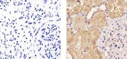
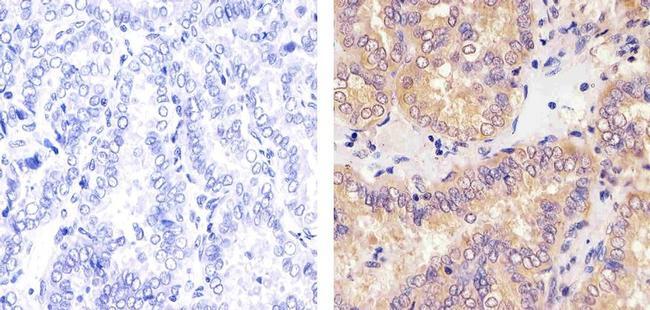
- Immunohistochemistry analysis of Cyclin D2 showing staining in the cytoplasm and nucleus of paraffin-embedded human thymus carcinoma (right) compared to a negative control without primary antibody (left). To expose target proteins, antigen retrieval was performed using 10mM sodium citrate (pH 6.0), microwaved for 8-15 min. Following antigen retrieval, tissues were blocked in 3% H2O2-methanol for 15 min at room temperature, washed with ddH2O and PBS, and then probed with a Cyclin D2 Antibody Mouse Monoclonal Antibody (Product # MA5-12731) diluted in 3% BSA-PBS at a dilution of 1:20 for 1 hour at 37°C in a humidified chamber. Tissues were washed extensively in PBST and detection was performed using an HRP-conjugated secondary antibody followed by colorimetric detection using a DAB kit. Tissues were counterstained with hematoxylin and dehydrated with ethanol and xylene to prep for mounting.
Supportive validation
- Submitted by
- Invitrogen Antibodies (provider)
- Main image
- Experimental details
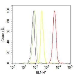
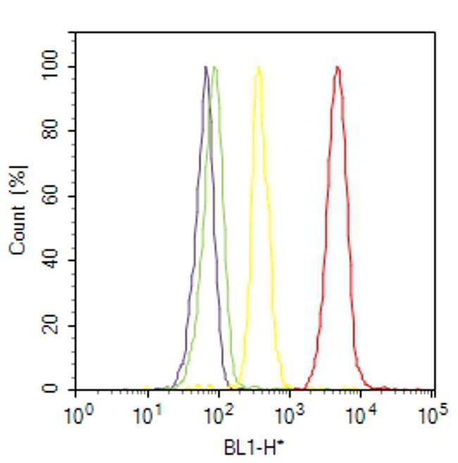
- Flow cytometry analysis of Cyclin D2 was done on NTERA-2 cells. Cells were fixed with 70% ethanol for 10 minutes, permeabilized with 0.25% Triton™ X-100 for 20 minutes, and blocked with 5% BSA for 30 minutes at room temperature. Cells were labeled with Cyclin D2 Mouse Monoclonal Antibody (MA512731, red histogram) or with mouse isotype control (yellow histogram) at 3-5 ug/million cells in 2.5% BSA. After incubation at room temperature for 2 hours, the cells were labeled with Alexa Fluor® 488 Rabbit Anti-Mouse Secondary Antibody (A11059) at a dilution of 1:400 for 30 minutes at room temperature. The representative 10,000 cells were acquired and analyzed for each sample using an Attune® Acoustic Focusing Cytometer. The purple histogram represents unstained control cells and the green histogram represents no-primary-antibody control.
Supportive validation
- Submitted by
- Invitrogen Antibodies (provider)
- Main image
- Experimental details
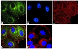
- Immunofluorescence analysis of Cyclin D2 was done on 70% confluent log phase MCF-7 cells. The cells were fixed with 4% paraformaldehyde for 10 minutes, permeabilized with 0.1% Triton™ X-100 for 10 minutes, and blocked with 1% BSA for 1 hour at room temperature. The cells were labeled with Cyclin D2 (DCS-3.1 + DCS-5.2) Mouse Monoclonal Antibody (Product # MA5-12731) at 2 µg/mL in 0.1% BSA and incubated for 3 hours at room temperature and then labeled with Goat anti-Mouse IgG (H+L) Superclonal™ Secondary Antibody, Alexa Fluor® 488 conjugate (Product # A28175) at a dilution of 1:2000 for 45 minutes at room temperature (Panel a: green). Nuclei (Panel b: blue) were stained with SlowFade® Gold Antifade Mountant with DAPI (Product # S36938). F-actin (Panel c: red) was stained with Alexa Fluor® 555 Rhodamine Phalloidin (Product # R415, 1:300). Panel d is a merged image showing cytoplasmic localization. Panel e is a no primary antibody control. The images were captured at 60X magnification.
