 Explore
 Validate
 Learn
Learn Western blot
Antibody data
- Antibody Data
- Antigen structure
- References [0]
- Comments [0]
- Validations
- Western blot [2]
- Immunocytochemistry [1]
- Immunohistochemistry [2]
- Other assay [1]
Submit
Validation data
Reference
Comment
Report error
- Product number
- MA5-27481 - Provider product page

- Provider
- Invitrogen Antibodies
- Product name
- IGF2BP3 Monoclonal Antibody (OTI7B10)
- Antibody type
- Monoclonal
- Antigen
- Recombinant full-length protein
- Reactivity
- Human, Mouse, Rat
- Host
- Mouse
- Isotype
- IgG
- Antibody clone number
- OTI7B10
- Vial size
- 100 µL
- Concentration
- 1 mg/mL
- Storage
- -20° C, Avoid Freeze/Thaw Cycles
No comments: Submit comment
Supportive validation
- Submitted by
- Invitrogen Antibodies (provider)
- Main image
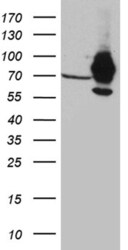
- Experimental details
- Western blot analysis of IGF2BP3 in HEK293T cells in untransfected (Left lane) and transfected (Right lane) samples using 5 µg per lane. The samples were separated by SDS-PAGE and probed with IGF2BP3 (Product # MA5-27481) monoclonal antibody.

- Submitted by
- Invitrogen Antibodies (provider)
- Main image
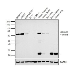
- Experimental details
- Western blot was performed using Anti-IGF2BP3 Mouse Monoclonal Antibody (Product # MA5-27481) and a 64 kDa band corresponding to IGF2BP3 was observed in cell lines and tissues tested except MCF7, SK-BR-3, Mouse Lung and Mouse Spleen as reported. Whole cell extracts (30 µg lysate) of SH-SY5Y (Lane 1), IMR-32 (Lane 2), MDA-MB-231 (Lane 3), MCF7 (Lane 4), SK-BR-3 (Lane 5), Mouse Placenta (Lane 6), Mouse Testis (Lane 7), Rat Testis (Lane 8), Mouse Spleen (Lane 9) and Mouse Lung (Lane 10) were electrophoresed using Novex® NuPAGE® 4-12% Bis-Tris Protein Gel (Product # NP0322BOX). Resolved proteins were then transferred onto a nitrocellulose membrane (Product # IB23001) by iBlot® 2 Dry Blotting System (Product # IB21001). The blot was probed with the primary antibody (1:1000 dilution) and detected by chemiluminescence with Goat anti-Mouse IgG (H+L) Superclonal™ Recombinant Secondary Antibody, HRP (Product # A28177, 1:4000 dilution) using the iBright FL 1000 (Product # A32752). Chemiluminescent detection was performed using Novex® ECL Chemiluminescent Substrate Reagent Kit (Product # WP20005). A 25 kDa band (*) corresponding to circulating tissue IgG, was observed in mouse tissues.
Supportive validation
- Submitted by
- Invitrogen Antibodies (provider)
- Main image

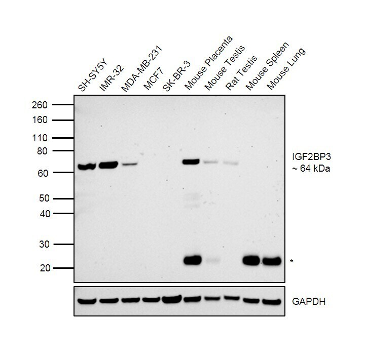
- Experimental details
- Immunofluorescence analysis of IGF2BP3 was performed using IMR-32 and MCF7 cells. The cells were fixed with 4% paraformaldehyde for 10 minutes, permeabilized with 0.1% Triton™ X-100 for 15 minutes, and blocked with 2% BSA for 1 hour at room temperature. The cells were labeled with IGF2BP3 Mouse Monoclonal Antibody (Product # MA5-27481) at 1:100 dilution in 0.1% BSA and incubated overnight at 4 degree and then labeled with Goat anti-Mouse IgG (H+L) Highly Cross-Adsorbed Secondary Antibody, Alexa Fluor Plus 488 (Product # A32723) at a dilution of 1:2000 for 45 minutes at room temperature (Panel a: green) in IMR-32 cells. Nuclei (Panel b: blue) were stained with ProLong™ Diamond Antifade Mountant with DAPI (Product # P36962). F-actin (Panel c: red) was stained with Rhodamine Phalloidin (Product # R415, 1:300). Panel d represents the merged image of IMR-32 cells, which is a positive model for IGF2BP3 expression showing a cytoplasmic and nuclear localization. Panel e represents the merged image ofMCF7 cells, that are null for IGF2BP3 protein expression. Panel f represents control cells with no primary antibody to assess background. The images were captured at 60X magnification.
Supportive validation
- Submitted by
- Invitrogen Antibodies (provider)
- Main image
- Experimental details
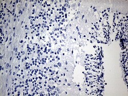
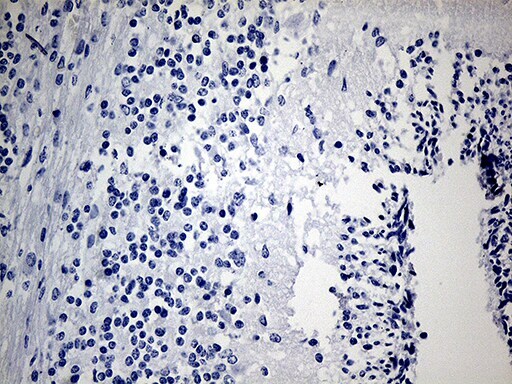
- Immunohistochemistry was performed on paraffin-embedded human embryonic cerebellum tissue. To expose target proteins, heat-induced epitope retrieval by 1mM EDTA in 10mM Tris buffer (pH8.5) at 120°C for 3 min. Following antigen retrieval, tissues were probed with a IGF2BP3 monoclonal antibody (Product # MA5-27481) at a dilution of 1:500.
- Submitted by
- Invitrogen Antibodies (provider)
- Main image
- Experimental details
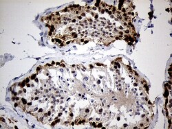
- Immunohistochemistry was performed on paraffin-embedded human testicle tissue. To expose target proteins, heat-induced epitope retrieval by 10mM citric buffer, pH6.0, 100°C for 10min. Following antigen retrieval, tissues were probed with a IGF2BP3 monoclonal antibody (Product # MA5-27481) at a dilution of 1:500.

Supportive validation
- Submitted by
- Invitrogen Antibodies (provider)
- Main image

- Experimental details
- RNA Immunoprecipitation (RIP) assay of endogenous IGF2BP3 protein using Anti-IGF2BP3 Antibody: RIP assay was performed using Anti-IGF2BP3 Monoclonal Antibody (Product # MA5-27481) 5 µg, on whole cell lysate from Hep G2 cells. Normal Mouse IgG was used as a negative IP control. RNA purified by RiboPure™ RNA Purification Kit (Product # AM1924) was analyzed by RT-PCR using the Power SYBR® Green RNA-to-CT™ 1-Step Kit (Product # 4389986) with the primers pairs over MYC exon1, IGF2 and 18S rRNA. Data is presented as fold enrichment of the antibody signal versus the negative control IgG using the comparative CT method.
