 Explore
 Validate
 Learn
Learn Western blot
 ELISA
ELISA Flow cytometry
Antibody data
- Antibody Data
- Antigen structure
- References [2]
- Comments [0]
- Validations
- Flow cytometry [1]
- Other assay [1]
Submit
Validation data
Reference
Comment
Report error
- Product number
- 39-8800 - Provider product page

- Provider
- Invitrogen Antibodies
- Product name
- IRF8 Monoclonal Antibody (ZI003)
- Antibody type
- Monoclonal
- Antigen
- Synthetic peptide
- Reactivity
- Human, Mouse
- Host
- Mouse
- Isotype
- IgG
- Antibody clone number
- ZI003
- Vial size
- 100 μg
- Concentration
- 0.5 mg/mL
- Storage
- -20°C
Submitted references Monocyte Subsets With High Osteoclastogenic Potential and Their Epigenetic Regulation Orchestrated by IRF8.
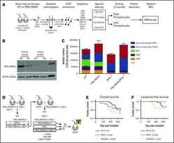
Identification of IRF8 as a potent tumor suppressor in murine acute promyelocytic leukemia.
Das A, Wang X, Kang J, Coulter A, Shetty AC, Bachu M, Brooks SR, Dell'Orso S, Foster BL, Fan X, Ozato K, Somerman MJ, Thumbigere-Math V
Journal of bone and mineral research : the official journal of the American Society for Bone and Mineral Research 2021 Jan;36(1):199-214
Journal of bone and mineral research : the official journal of the American Society for Bone and Mineral Research 2021 Jan;36(1):199-214
Identification of IRF8 as a potent tumor suppressor in murine acute promyelocytic leukemia.
Gaillard C, Surianarayanan S, Bentley T, Warr MR, Fitch B, Geng H, Passegué E, de Thé H, Kogan SC
Blood advances 2018 Oct 9;2(19):2462-2466
Blood advances 2018 Oct 9;2(19):2462-2466
No comments: Submit comment
Supportive validation
- Submitted by
- Invitrogen Antibodies (provider)
- Main image
- Experimental details
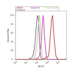
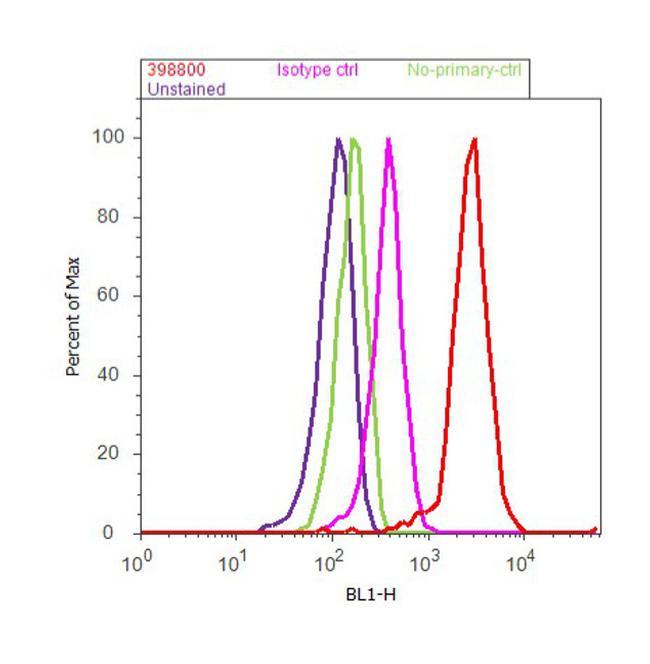
- Flow cytometry analysis of IRF8 was done on U-937 cells. Cells were fixed with 70% ethanol for 10 minutes, permeabilized with 0.25% Triton™ X-100 for 20 minutes, and blocked with 5% BSA for 30 minutes at room temperature. Cells were labeled with IRF8 Mouse Monoclonal Antibody (398800, red histogram) or with mouse isotype control (pink histogram) at 3-5 ug/million cells in 2.5% BSA. After incubation at room temperature for 2 hours, the cells were labeled with Alexa Fluor® 488 Rabbit Anti-Mouse Secondary Antibody (A11059) at a dilution of 1:400 for 30 minutes at room temperature. The representative 10,000 cells were acquired and analyzed for each sample using an Attune® Acoustic Focusing Cytometer. The purple histogram represents unstained control cells and the green histogram represents no-primary-antibody control..
Supportive validation
- Submitted by
- Invitrogen Antibodies (provider)
- Main image
- Experimental details
- NULL
