Explore
 Validate
 Learn
Learn Western blot
Antibody data
- Antibody Data
- Antigen structure
- References [1]
- Comments [0]
- Validations
- Western blot [2]
- Immunocytochemistry [1]
Submit
Validation data
Reference
Comment
Report error
- Product number
- 702323 - Provider product page

- Provider
- Invitrogen Antibodies
- Product name
- IRF8 Recombinant Rabbit Monoclonal Antibody (24H13L20)
- Antibody type
- Monoclonal
- Antigen
- Synthetic peptide
- Reactivity
- Human
- Host
- Rabbit
- Isotype
- IgG
- Antibody clone number
- 24H13L20
- Vial size
- 100 µg
- Concentration
- 0.5 mg/mL
- Storage
- Store at 4°C short term. For long term storage, store at -20°C, avoiding freeze/thaw cycles.
Submitted references Perturbation of astroglial Slc38 glutamine transporters by NH(4)(+) contributes to neurophysiologic manifestations in acute liver failure.
Hamdani EH, Popek M, Frontczak-Baniewicz M, Utheim TP, Albrecht J, Zielińska M, Chaudhry FA
FASEB journal : official publication of the Federation of American Societies for Experimental Biology 2021 Jul;35(7):e21588
FASEB journal : official publication of the Federation of American Societies for Experimental Biology 2021 Jul;35(7):e21588
No comments: Submit comment
Supportive validation
- Submitted by
- Invitrogen Antibodies (provider)
- Main image
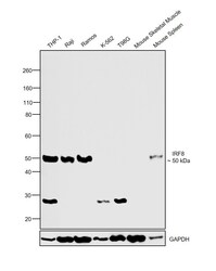
- Experimental details
- Western blot was performed using Anti-IRF8 Recombinant Rabbit Monoclonal Antibody (24H13L20)(Product # 702323) and a 50kDa band corresponding to IRF8 was observed across cell lines and tissue extract tested except K-562, T98G and Mouse Skeletal Muscle which are reported to be negative. Nuclear enriched extracts (30 µg lysate) of THP-1 (Lane 1), Raji (Lane 2), Ramos (Lane 3), K-562 (Lane 4), T98G (Lane 5) and tissue extracts of Mouse Skeletal Muscle (Lane 6) and Mouse Spleen (Lane 7) were electrophoresed using NuPAGE™ 4-12% Bis-Tris Protein Gel (Product # NP0322BOX). Resolved proteins were then transferred onto a Nitrocellulose membrane (Product # IB23001) by iBlot® 2 Dry Blotting System (Product # IB21001). The blot was probed with the primary antibody (1 ug/ml) and detected by chemiluminescence with Goat anti-Rabbit IgG (H+L) Superclonal™ Recombinant Secondary Antibody, HRP (Product # A27036,1:4000 dilution) using the iBright FL 1000 (Product # A32752). Chemiluminescent detection was performed using Novex® ECL Chemiluminescent Substrate Reagent Kit (Product # WP20005).
- Submitted by
- Invitrogen Antibodies (provider)
- Main image
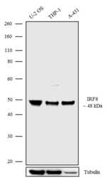
- Experimental details
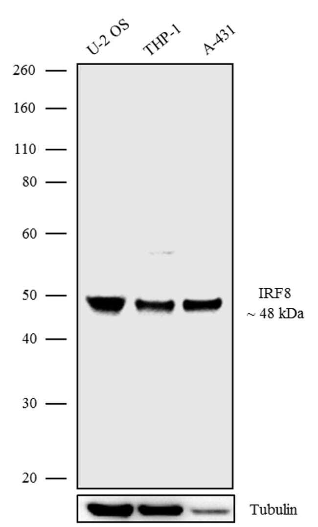
- Western blot analysis was performed on Whole cell extracts (30 µg lysate) of U-2 OS (Lane 1), THP-1 (Lane 2) and A-431 (Lane 3). The blots were probed with Anti-IRF8 Recombinant Rabbit Monoclonal Antibody (Product # 702323, 1-2 µg/mL) and detected by chemiluminescence using Goat anti-Rabbit IgG (H+L) Superclonal Secondary Antibody, HRP conjugate (Product # A27036, 0.4 µg/mL, 1:2500 dilution). A 48 kDa band corresponding to IRF8 was observed across the cell lines tested. Known quantity of protein samples were electrophoresed using Novex®NuPAGE®4-12% Bis-Tris gel (Product # NP0321BOX), XCell SureLock Electrophoresis System (Product # EI0002) and Novex® Sharp Pre-Stained Protein Standard (Product # LC5800). Resolved proteins were then transferred onto a nitrocellulose membrane with iBlot® Dry Blotting System (Product # IB21001). The membrane was probed with the relevant primary and secondary Antibody following blocking with 5% skimmed milk. Chemiluminescent detection was performed using Pierce™ ECL Western blotting Substrate (Product # 32106).
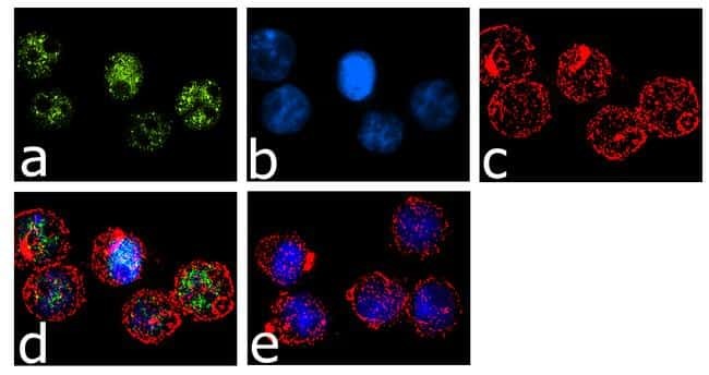
Supportive validation
- Submitted by
- Invitrogen Antibodies (provider)
- Main image

- Experimental details
- For immunofluorescence analysis, THP1 cells were fixed and permeabilized for detection of endogenous IRF8 using Anti- IRF8 Recombinant Rabbit Monoclonal Antibody (Product # 702323, 2 µg/mL) and labeled with Goat anti-Rabbit IgG (H+L) Superclonal Secondary Antibody, Alexa Fluor® 488 conjugate (Product # A27034, 1:2000). Panel a) shows representative cells that were stained for detection and localization of IRF8 protein (green), Panel b) is stained for nuclei (blue) using SlowFade® Gold Antifade Mountant with DAPI (Product # S36938). Panel c) represents cytoskeletal F-actin staining using Alexa Fluor® 555 Rhodamine Phalloidin (Product # R415, 1:300). Panel d) is a composite image of Panels a, b and c clearly demonstrating nuclear localization of IRF8. Panel e) represents control cells with no primary antibody to assess background. The images were captured at 60X magnification.