 Explore
 Validate
 Learn
Learn Western blot
Antibody data
- Antibody Data
- Antigen structure
- References [12]
- Comments [0]
- Validations
- Western blot [3]
- Immunohistochemistry [2]
Submit
Validation data
Reference
Comment
Report error
- Product number
- AF1757 - Provider product page

- Provider
- Novus Biologicals
- Product name
- Goat Polyclonal Lipocalin-2/NGAL Antibody
- Antibody type
- Polyclonal
- Description
- Antigen Affinity-purified. Detects human Lipocalin-2/NGAL in direct ELISAs and Western blots.
- Reactivity
- Human, Mouse, Rat
- Host
- Goat
- Conjugate
- Unconjugated
- Isotype
- IgG
- Vial size
- 100 ug
- Concentration
- LYOPH
- Storage
- Use a manual defrost freezer and avoid repeated freeze-thaw cycles. 12 months from date of receipt, -20 to -70 degreesC as supplied. 1 month, 2 to 8 degreesC under sterile conditions after reconstitution. 6 months, -20 to -70 degreesC under sterile conditions after reconstitution.
Submitted references Lipocalin-2 Regulates Epidermal Growth Factor Receptor Intracellular Trafficking.
Novel aroylated phenylenediamine compounds enhance antimicrobial defense and maintain airway epithelial barrier integrity.
CLEC3A, MMP7, and LCN2 as novel markers for predicting recurrence in resected G1 and G2 pancreatic neuroendocrine tumors.
Unexpected kidney-restricted role for IL-17 receptor signaling in defense against systemic Candida albicans infection.
Cilostazol attenuates kainic acid-induced hippocampal cell death.
Glycine mitigates renal oxidative stress by suppressing Nox4 expression in rats with streptozotocin-induced diabetes.
Metformin alleviates nickel-induced autophagy and apoptosis via inhibition of hexokinase-2, activating lipocalin-2, in human bronchial epithelial cells.
Expression of uterine lipocalin 2 and its receptor during early- to mid-pregnancy period in mares.
The adipokine lipocalin-2 in the context of the osteoarthritic osteochondral junction.
Lipocalin2 promotes invasion, tumorigenicity and gemcitabine resistance in pancreatic ductal adenocarcinoma.
Cellular settings mediating Src Substrate switching between focal adhesion kinase tyrosine 861 and CUB-domain-containing protein 1 (CDCP1) tyrosine 734.
Urinary neutrophil gelatinase-associated lipocalin levels reflect damage to glomeruli, proximal tubules, and distal nephrons.
Yammine L, Zablocki A, Baron W, Terzi F, Gallazzini M
Cell reports 2019 Nov 12;29(7):2067-2077.e6
Cell reports 2019 Nov 12;29(7):2067-2077.e6
Novel aroylated phenylenediamine compounds enhance antimicrobial defense and maintain airway epithelial barrier integrity.
Myszor IT, Parveen Z, Ottosson H, Bergman P, Agerberth B, Strömberg R, Gudmundsson GH
Scientific reports 2019 May 8;9(1):7114
Scientific reports 2019 May 8;9(1):7114
CLEC3A, MMP7, and LCN2 as novel markers for predicting recurrence in resected G1 and G2 pancreatic neuroendocrine tumors.
Miki M, Oono T, Fujimori N, Takaoka T, Kawabe K, Miyasaka Y, Ohtsuka T, Saito D, Nakamura M, Ohkawa Y, Oda Y, Suyama M, Ito T, Ogawa Y
Cancer medicine 2019 Jul;8(8):3748-3760
Cancer medicine 2019 Jul;8(8):3748-3760
Unexpected kidney-restricted role for IL-17 receptor signaling in defense against systemic Candida albicans infection.
Ramani K, Jawale CV, Verma AH, Coleman BM, Kolls JK, Biswas PS
JCI insight 2018 May 3;3(9)
JCI insight 2018 May 3;3(9)
Cilostazol attenuates kainic acid-induced hippocampal cell death.
Park YS, Jin Z, Jeong EA, Yi CO, Lee JY, Park IS, Roh GS
The Korean journal of physiology & pharmacology : official journal of the Korean Physiological Society and the Korean Society of Pharmacology 2018 Jan;22(1):63-70
The Korean journal of physiology & pharmacology : official journal of the Korean Physiological Society and the Korean Society of Pharmacology 2018 Jan;22(1):63-70
Glycine mitigates renal oxidative stress by suppressing Nox4 expression in rats with streptozotocin-induced diabetes.
Wang Z, Zhang J, Wang L, Li W, Chen L, Li J, Zhao D, Zhang H, Guo X
Journal of pharmacological sciences 2018 Aug;137(4):387-394
Journal of pharmacological sciences 2018 Aug;137(4):387-394
Metformin alleviates nickel-induced autophagy and apoptosis via inhibition of hexokinase-2, activating lipocalin-2, in human bronchial epithelial cells.
Kang YT, Hsu WC, Wu CH, Hsin IL, Wu PR, Yeh KT, Ko JL
Oncotarget 2017 Dec 1;8(62):105536-105552
Oncotarget 2017 Dec 1;8(62):105536-105552
Expression of uterine lipocalin 2 and its receptor during early- to mid-pregnancy period in mares.
Haneda S, Nagaoka K, Nambo Y, Kikuchi M, Nakano Y, Li J, Matsui M, Miyake YI, Imakawa K
The Journal of reproduction and development 2017 Apr 21;63(2):127-133
The Journal of reproduction and development 2017 Apr 21;63(2):127-133
The adipokine lipocalin-2 in the context of the osteoarthritic osteochondral junction.
Villalvilla A, García-Martín A, Largo R, Gualillo O, Herrero-Beaumont G, Gómez R
Scientific reports 2016 Jul 7;6:29243
Scientific reports 2016 Jul 7;6:29243
Lipocalin2 promotes invasion, tumorigenicity and gemcitabine resistance in pancreatic ductal adenocarcinoma.
Leung L, Radulovich N, Zhu CQ, Organ S, Bandarchi B, Pintilie M, To C, Panchal D, Tsao MS
PloS one 2012;7(10):e46677
PloS one 2012;7(10):e46677
Cellular settings mediating Src Substrate switching between focal adhesion kinase tyrosine 861 and CUB-domain-containing protein 1 (CDCP1) tyrosine 734.
Wortmann A, He Y, Christensen ME, Linn M, Lumley JW, Pollock PM, Waterhouse NJ, Hooper JD
The Journal of biological chemistry 2011 Dec 9;286(49):42303-42315
The Journal of biological chemistry 2011 Dec 9;286(49):42303-42315
Urinary neutrophil gelatinase-associated lipocalin levels reflect damage to glomeruli, proximal tubules, and distal nephrons.
Kuwabara T, Mori K, Mukoyama M, Kasahara M, Yokoi H, Saito Y, Yoshioka T, Ogawa Y, Imamaki H, Kusakabe T, Ebihara K, Omata M, Satoh N, Sugawara A, Barasch J, Nakao K
Kidney international 2009 Feb;75(3):285-94
Kidney international 2009 Feb;75(3):285-94
No comments: Submit comment
Supportive validation
- Submitted by
- Novus Biologicals (provider)
- Main image
- Experimental details
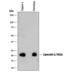
- Detection of Human Lipocalin-2/NGAL by Western Blot. Western blot shows lysates of Capan-1 human pancreatic adenocarcinoma cell line and human pancreas tissue. PVDF membrane was probed with 0.2 µg/mL of Goat Anti-Human/Mouse/Rat Lipocalin-2/NGAL Antigen Affinity-purified Polyclonal Antibody (Catalog # AF1757) followed by HRP-conjugated Anti-Goat IgG Secondary Antibody (Catalog # HAF109). A specific band was detected for Lipocalin-2/NGAL at approximately 22 kDa (as indicated). This experiment was conducted under reducing conditions and using Immunoblot Buffer Group 1.

- Submitted by
- Novus Biologicals (provider)
- Main image

- Experimental details
- Detection of Mouse and Rat Lipocalin-2/NGAL by Western Blot. Western blot shows lysates of mouse and rat bone marrow. PVDF membrane was probed with 0.2 µg/mL of Goat Anti-Human/Mouse/Rat Lipocalin-2/NGAL Antigen Affinity-purified Polyclonal Antibody (Catalog # AF1757) followed by HRP-conjugated Anti-Goat IgG Secondary Antibody (Catalog # HAF017). A specific band was detected for Lipocalin-2/NGAL at approximately 22 kDa (as indicated). This experiment was conducted under reducing conditions and using Immunoblot Buffer Group 1.

- Submitted by
- Novus Biologicals (provider)
- Main image
- Experimental details
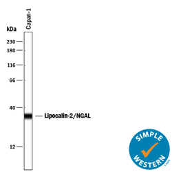
- Detection of Human Lipocalin-2/NGAL by Simple WesternTM. Simple Western lane view shows lysates of Capan-1 human pancreatic adenocarcinoma cell line, loaded at 0.2 mg/mL. A specific band was detected for Lipocalin-2/NGAL at approximately 34 kDa (as indicated) using 2 µg/mL of Goat Anti-Human/Mouse/Rat Lipocalin-2/NGAL Antigen Affinity-purified Polyclonal Antibody (Catalog # AF1757) followed by 1:50 dilution of HRP-conjugated Anti-Goat IgG Secondary Antibody (Catalog # HAF109). This experiment was conducted under reducing conditions and using the 12-230 kDa separation system.

Supportive validation
- Submitted by
- Novus Biologicals (provider)
- Main image
- Experimental details
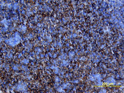
- Lipocalin-2/NGAL in Human Pancreatic Cancer Tissue. Lipocalin-2/NGAL was detected in immersion fixed paraffin-embedded sections of human pancreatic cancer tissue using Goat Anti-Human/Mouse/ Rat Lipocalin-2/NGAL Antigen Affinity-purified Polyclonal Antibody (Catalog # AF1757) at 15 µg/mL overnight at 4 °C. Tissue was stained using the Anti-Goat HRP-DAB Cell & Tissue Staining Kit (brown; Catalog # CTS008) and counter-stained with hematoxylin (blue). View our protocol for Chromogenic IHC Staining of Paraffin-embedded Tissue Sections.

- Submitted by
- Novus Biologicals (provider)
- Main image

- Experimental details
- Lipocalin-2/NGAL in Human Pancreatic Cancer Tissue. Lipocalin-2/NGAL was detected in immersion fixed paraffin-embedded sections of human pancreatic cancer tissue using Goat Anti-Human/Mouse/Rat Lipocalin-2/NGAL Antigen Affinity-purified Polyclonal Antibody (Catalog # AF1757) at 15 µg/mL overnight at 4 °C. Tissue was stained using the Anti-Goat HRP-DAB Cell & Tissue Staining Kit (brown; Catalog # CTS008) and counterstained with hematoxylin (blue). Lower panel shows a lack of labeling if primary antibodies are omitted and tissue is stained only with secondary antibody followed by incubation with detection reagents. View our protocol for Chromogenic IHC Staining of Paraffin-embedded Tissue Sections.
