 Explore
 Validate
 Learn
Learn Flow cytometry
Antibody data
- Antibody Data
- Antigen structure
- References [1]
- Comments [0]
- Validations
- Flow cytometry [1]
Submit
Validation data
Reference
Comment
Report error
- Product number
- 12-9176-42 - Provider product page

- Provider
- Invitrogen Antibodies
- Product name
- Survivin Monoclonal Antibody (STLALYV), PE, eBioscience™
- Antibody type
- Monoclonal
- Antigen
- Other
- Description
- Description: This STLALYV monoclonal antibody recognizes human survivin (also known as Birc5). Survivin is a member of the inhibitor of apoptosis (IAP) family that also includes XIAP, c-IAP1, c-IAP2, and NAIP. Survivin functions to inhibit caspase activation, thereby leading to negative regulation of apoptosis. Survivin also plays a critical role in mitosis and cytokinesis where it is expressed only during G2/M and localizes to the mitotic spindle and is found at the cleavage furrow/midplate. Thus, survivin is highly expressed in tumors and in fetal tissue, but is absent in terminally-differentiated cells and non-proliferating cells. Deletion of survivin in developing thymocytes as well as in mature T cells showed that survivin is critical for cell division in lymphoid cells but not for inhibition of survival; the same is true for B lymphocytes. Applications Reported: This STLALYV antibody has been reported for use in intracellular staining followed by flow cytometric analysis. Applications Tested: This STLALYV antibody has been pre-titrated and tested by intracellular staining followed by flow cytometric analysis of stimulated normal human peripheral blood cells using the Foxp3/Transcription Factor Staining Buffer Set (Product # 00-5523-00) and protocol. Please refer to Best Protocols: Protocol B: One step protocol for (nuclear) intracellular proteins located under the Resources Tab online. This can be used at 5 µL (0.5 µg) per test. A test is defined as the amount (µg) of antibody that will stain a cell sample in a final volume of 100 µL. Cell number should be determined empirically but can range from 10^5 to 10^8 cells/test. Excitation: 488-561 nm; Emission: 578 nm; Laser: Blue Laser, Green Laser, Yellow-Green Laser. Filtration: 0.2 µm post-manufacturing filtered.
- Reactivity
- Human
- Host
- Mouse
- Conjugate
- Yellow dye
- Isotype
- IgG
- Antibody clone number
- STLALYV
- Vial size
- 100 Tests
- Concentration
- 5 µL/Test
- Storage
- 4° C, store in dark, DO NOT FREEZE!
Submitted references Umbilical cord-derived mesenchymal stem cells inhibit growth and promote apoptosis of HepG2 cells.
Tang YM, Bao WM, Yang JH, Ma LK, Yang J, Xu Y, Yang LH, Sha F, Xu ZY, Wu HM, Zhou W, Li Y, Li YH
Molecular medicine reports 2016 Sep;14(3):2717-24
Molecular medicine reports 2016 Sep;14(3):2717-24
No comments: Submit comment
Supportive validation
- Submitted by
- Invitrogen Antibodies (provider)
- Main image
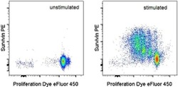
- Experimental details
- Intracellular staining of Cell Proliferation Dye eFluor® 450 (Product # 65-0842-85)-labeled normal human peripheral cells that were unstimulated (left) or Con A (Product # 00-4978-03)-stimulated (right) with Anti-Human Survivin PE using the Foxp3/Transcription Factor Staining Buffer Set (Product # 00-5523-00) and protocol. Total viable cells, as determined by Fixable Viability Dye eFluor® 506 (Product # 65-0866-14), were used for analysis.
- Conjugate
- Yellow dye
