Explore
 Validate
 Learn
Learn Immunocytochemistry
Antibody data
- Antibody Data
- Antigen structure
- References [3]
- Comments [0]
- Validations
- Immunocytochemistry [3]
- Immunohistochemistry [1]
Submit
Validation data
Reference
Comment
Report error
- Product number
- 14-9176-82 - Provider product page

- Provider
- Invitrogen Antibodies
- Product name
- Survivin Monoclonal Antibody (STLALYV), eBioscience™
- Antibody type
- Monoclonal
- Antigen
- Other
- Description
- Description: This STLALYV monoclonal antibody recognizes human survivin (also known as Birc5). Survivin is a member of the inhibitor of apoptosis (IAP) family that also includes XIAP, c-IAP1, c-IAP2, and NAIP. Survivin functions to inhibit caspase activation, thereby leading to negative regulation of apoptosis. Survivin also plays a critical role in mitosis and cytokinesis where it is expressed only during G2/M and localizes to the mitotic spindle and is found at the cleavage furrow/midplate. Thus, survivin is highly expressed in tumors and in fetal tissue, but is absent in terminally-differentiated cells and non-proliferating cells. Deletion of survivin in developing thymocytes as well as in mature T cells showed that survivin is critical for cell division in lymphoid cells but not for inhibition of survival; the same is true for B lymphocytes. Applications Reported: This STLALYV antibody has been reported for use in immunohistochemical staining of formalin-fixed paraffin embedded tissue sections, microscopy, and immunocytochemistry. (Fluorochrome-conjugated STLALYV is recommended for use in intracellular flow cytometry.). Applications Tested: This STLALYV antibody has been tested by immunocytochemistry of formaldehyde-fixed and permeabilized cells and can be used at less than or equal to 5 µg/mL. This STLALYV antibody has also been tested by immunohistochemistry of formalin-fixed paraffin embedded tissue using high pH antigen retrieval and can be used at less than or equal to 5 µg/mL. It is recommended that the antibody be carefully titrated for optimal performance in the assay of interest. Purity: Greater than 90%, as determined by SDS-PAGE. Aggregation: Less than 10%, as determined by HPLC. Filtration: 0.2 µm post-manufacturing filtered.
- Reactivity
- Human
- Host
- Mouse
- Isotype
- IgG
- Antibody clone number
- STLALYV
- Vial size
- 100 µg
- Concentration
- 0.5 mg/mL
- Storage
- 4° C
Submitted references Treat cancers by targeting survivin: just a dream or future reality?
Sustained survivin expression from OX40 costimulatory signals drives T cell clonal expansion.
Survivin loss in thymocytes triggers p53-mediated growth arrest and p53-independent cell death.
Coumar MS, Tsai FY, Kanwar JR, Sarvagalla S, Cheung CH
Cancer treatment reviews 2013 Nov;39(7):802-11
Cancer treatment reviews 2013 Nov;39(7):802-11
Sustained survivin expression from OX40 costimulatory signals drives T cell clonal expansion.
Song J, So T, Cheng M, Tang X, Croft M
Immunity 2005 May;22(5):621-31
Immunity 2005 May;22(5):621-31
Survivin loss in thymocytes triggers p53-mediated growth arrest and p53-independent cell death.
Okada H, Bakal C, Shahinian A, Elia A, Wakeham A, Suh WK, Duncan GS, Ciofani M, Rottapel R, Zúñiga-Pflücker JC, Mak TW
The Journal of experimental medicine 2004 Feb 2;199(3):399-410
The Journal of experimental medicine 2004 Feb 2;199(3):399-410
No comments: Submit comment
Supportive validation
- Submitted by
- Invitrogen Antibodies (provider)
- Main image

- Experimental details
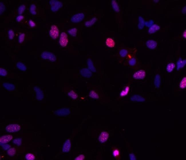
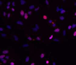
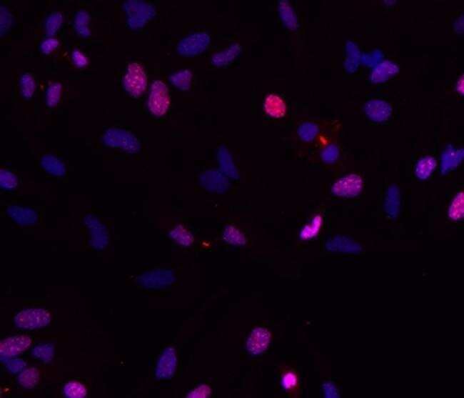
- Immunocytochemistry of fixed and permeabilized HeLa cells using 5 µg/mL Anti-Human Survivin Purified, followed by F (ab')2 Anti-Mouse IgG eFluor® 570.Nuclei are stained with DAPI.
- Submitted by
- Invitrogen Antibodies (provider)
- Main image
- Experimental details
- Immunocytochemistry of fixed and permeabilized HeLa cells using 5 µg/mL Anti-Human Survivin Purified, followed by F (ab')2 Anti-Mouse IgG eFluor® 570.Nuclei are stained with DAPI.
- Submitted by
- Invitrogen Antibodies (provider)
- Main image

- Experimental details
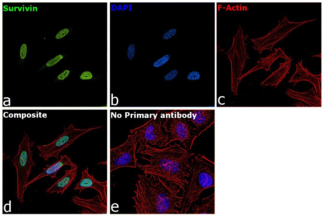
- Immunofluorescence analysis of Survivin was performed using 70% confluent log phase HeLa cells. The cells were fixed with 4% paraformaldehyde for 10 minutes, permeabilized with 0.1% Triton™ X-100 for 15 minutes, and blocked with 2% BSA for 45 minutes at room temperature. The cells were labeled with Survivin Monoclonal Antibody (STLALYV), eBioscience™ (Product # 14-9176-82) at 5 µg/mL in 0.1% BSA, incubated at 4 degree celsius overnight and then labeled with Goat anti-Mouse IgG (H+L) Superclonal™ Recombinant Secondary Antibody, Alexa Fluor® 488 conjugate (Product # A28175), (1:2000 dilution), for 45 minutes at room temperature (Panel a: Green). Nuclei (Panel b:Blue) were stained with ProLong™ Diamond Antifade Mountant with DAPI (Product # P36962). F-actin (Panel c: Red) was stained with Rhodamine Phalloidin (Product # R415, 1:300). Panel d represents the merged image showing Nuclear localization. Panel e represents control cells with no primary antibody to assess background. The images were captured at 60X magnification.
Supportive validation
- Submitted by
- Invitrogen Antibodies (provider)
- Main image
- Experimental details
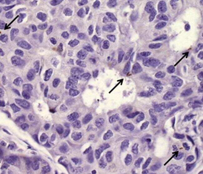
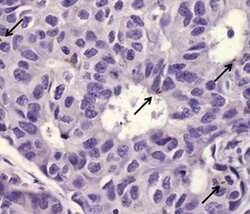
- Immunohistochemistry of formalin-fixed paraffin embedded human breast cancer tissue using 5 µg/mL Anti-Human Survivin Purified, followed by Anti-Mouse IgG Biotin, Streptavidin HRP and DAB visualization (arrows, right).Nuclei are counterstained with hematoxylin.