Explore
 Validate
 Learn
Learn Immunocytochemistry
Antibody data
- Antibody Data
- Antigen structure
- References [16]
- Comments [0]
- Validations
- Immunocytochemistry [3]
- Immunohistochemistry [3]
Submit
Validation data
Reference
Comment
Report error
- Product number
- MA5-11680 - Provider product page

- Provider
- Invitrogen Antibodies
- Product name
- Survivin Monoclonal Antibody (8E2)
- Antibody type
- Monoclonal
- Antigen
- Recombinant full-length protein
- Description
- MA5-11680 targets Survivin in ICC/IF and IHC (P) applications and shows reactivity with Human and Rat samples. The MA5-11680 immunogen is full length recombinant human survivin protein.
- Reactivity
- Human, Rat
- Host
- Mouse
- Isotype
- IgG
- Antibody clone number
- 8E2
- Vial size
- 500 µL
- Concentration
- 0.2 mg/mL
- Storage
- 4° C
Submitted references Arsenic trioxide treatment of rabbit liver VX-2 carcinoma via hepatic arterial cannulation-induced apoptosis and decreased levels of survivin in the tumor tissue.
Prognostication of soft tissue sarcomas based on chromosome 17q gene and protein status: evaluation of TOP2A, HER-2/neu, and survivin.
Ex vivo enrichment of circulating anti-tumor T cells from both cutaneous and ocular melanoma patients: clinical implications for adoptive cell transfer therapy.
Structure-specific recognition protein 1 facilitates microtubule growth and bundling required for mitosis.
Immunobiological characterization of cancer stem cells isolated from glioblastoma patients.
Norcantharidin inhibits growth of human gallbladder carcinoma xenografted tumors in nude mice by inducing apoptosis and blocking the cell cycle in vivo.
Survivin expression quantified by Image Pro-Plus compared with visual assessment.
Significance of an exon 2 G4C14-to-A4T14 polymorphism in the p73 gene on survival in rectal cancer patients with or without preoperative radiotherapy.
p53-dependent antiproliferative and antitumor effect of novel alkyl series of diorganotin(IV) compounds.
Deoxycholic acid promotes the growth of colonic aberrant crypt foci.
Expression of S100A4 and Met: potential predictors for metastasis and survival in early-stage breast cancer.
Survivin expression is a prognostic marker in pancreatic cancer patients.
Inhibitor of apoptosis protein (IAP) survivin is upregulated by oncogenic c-H-Ras.
Expression of the inhibitor of apoptosis protein survivin in benign meningiomas.
Expression of the inhibitor of apoptosis protein survivin in benign meningiomas.
Expression of survivin in primary glioblastomas.
Li H, Gong J, Jiang X, Shao H
Croatian medical journal 2013 Feb;54(1):12-6
Croatian medical journal 2013 Feb;54(1):12-6
Prognostication of soft tissue sarcomas based on chromosome 17q gene and protein status: evaluation of TOP2A, HER-2/neu, and survivin.
da Cunha IW, De Brot L, Carvalho KC, Rocha RM, Fregnani JH, Falzoni R, Ferreira Fde O, Aguiar S Jr, Lopes A, Muto NH, Reis LF, Soares FA, Vassallo J
Annals of surgical oncology 2012 Jun;19(6):1790-9
Annals of surgical oncology 2012 Jun;19(6):1790-9
Ex vivo enrichment of circulating anti-tumor T cells from both cutaneous and ocular melanoma patients: clinical implications for adoptive cell transfer therapy.
Mazzarella T, Cambiaghi V, Rizzo N, Pilla L, Parolini D, Orsenigo E, Colucci A, Modorati G, Doglioni C, Parmiani G, Maccalli C
Cancer immunology, immunotherapy : CII 2012 Aug;61(8):1169-82
Cancer immunology, immunotherapy : CII 2012 Aug;61(8):1169-82
Structure-specific recognition protein 1 facilitates microtubule growth and bundling required for mitosis.
Zeng SX, Li Y, Jin Y, Zhang Q, Keller DM, McQuaw CM, Barklis E, Stone S, Hoatlin M, Zhao Y, Lu H
Molecular and cellular biology 2010 Feb;30(4):935-47
Molecular and cellular biology 2010 Feb;30(4):935-47
Immunobiological characterization of cancer stem cells isolated from glioblastoma patients.
Di Tomaso T, Mazzoleni S, Wang E, Sovena G, Clavenna D, Franzin A, Mortini P, Ferrone S, Doglioni C, Marincola FM, Galli R, Parmiani G, Maccalli C
Clinical cancer research : an official journal of the American Association for Cancer Research 2010 Feb 1;16(3):800-13
Clinical cancer research : an official journal of the American Association for Cancer Research 2010 Feb 1;16(3):800-13
Norcantharidin inhibits growth of human gallbladder carcinoma xenografted tumors in nude mice by inducing apoptosis and blocking the cell cycle in vivo.
Fan YZ, Zhao ZM, Fu JY, Chen CQ, Sun W
Hepatobiliary & pancreatic diseases international : HBPD INT 2010 Aug;9(4):414-22
Hepatobiliary & pancreatic diseases international : HBPD INT 2010 Aug;9(4):414-22
Survivin expression quantified by Image Pro-Plus compared with visual assessment.
Wang CJ, Zhou ZG, Holmqvist A, Zhang H, Li Y, Adell G, Sun XF
Applied immunohistochemistry & molecular morphology : AIMM 2009 Dec;17(6):530-5
Applied immunohistochemistry & molecular morphology : AIMM 2009 Dec;17(6):530-5
Significance of an exon 2 G4C14-to-A4T14 polymorphism in the p73 gene on survival in rectal cancer patients with or without preoperative radiotherapy.
Lööf J, Pfeifer D, Adell G, Sun XF
Radiotherapy and oncology : journal of the European Society for Therapeutic Radiology and Oncology 2009 Aug;92(2):215-20
Radiotherapy and oncology : journal of the European Society for Therapeutic Radiology and Oncology 2009 Aug;92(2):215-20
p53-dependent antiproliferative and antitumor effect of novel alkyl series of diorganotin(IV) compounds.
Koch B, Basu Baul TS, Chatterjee A
Investigational new drugs 2009 Aug;27(4):319-26
Investigational new drugs 2009 Aug;27(4):319-26
Deoxycholic acid promotes the growth of colonic aberrant crypt foci.
Flynn C, Montrose DC, Swank DL, Nakanishi M, Ilsley JN, Rosenberg DW
Molecular carcinogenesis 2007 Jan;46(1):60-70
Molecular carcinogenesis 2007 Jan;46(1):60-70
Expression of S100A4 and Met: potential predictors for metastasis and survival in early-stage breast cancer.
Lee WY, Su WC, Lin PW, Guo HR, Chang TW, Chen HH
Oncology 2004;66(6):429-38
Oncology 2004;66(6):429-38
Survivin expression is a prognostic marker in pancreatic cancer patients.
Kami K, Doi R, Koizumi M, Toyoda E, Mori T, Ito D, Fujimoto K, Wada M, Miyatake S, Imamura M
Surgery 2004 Aug;136(2):443-8
Surgery 2004 Aug;136(2):443-8
Inhibitor of apoptosis protein (IAP) survivin is upregulated by oncogenic c-H-Ras.
Sommer KW, Schamberger CJ, Schmidt GE, Sasgary S, Cerni C
Oncogene 2003 Jul 3;22(27):4266-80
Oncogene 2003 Jul 3;22(27):4266-80
Expression of the inhibitor of apoptosis protein survivin in benign meningiomas.
Das A, Tan WL, Smith DR
Cancer letters 2003 Apr 25;193(2):217-23
Cancer letters 2003 Apr 25;193(2):217-23
Expression of the inhibitor of apoptosis protein survivin in benign meningiomas.
Das A, Tan WL, Smith DR
Cancer letters 2003 Apr 25;193(2):217-23
Cancer letters 2003 Apr 25;193(2):217-23
Expression of survivin in primary glioblastomas.
Das A, Tan WL, Teo J, Smith DR
Journal of cancer research and clinical oncology 2002 Jun;128(6):302-6
Journal of cancer research and clinical oncology 2002 Jun;128(6):302-6
No comments: Submit comment
Supportive validation
- Submitted by
- Invitrogen Antibodies (provider)
- Main image

- Experimental details
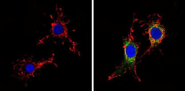
- Immunofluorescent analysis of Survivin (green) showing staining in the cytoplasm of A431 cells (right) compared to a negative control without primary antibody (left). Formalin-fixed cells were permeabilized with 0.1% Triton X-100 in TBS for 5-10 minutes and blocked with 3% BSA-PBS for 30 minutes at room temperature. Cells were probed with a Survivin monoclonal antibody (Product # MA5-11680) in 3% BSA-PBS at a dilution of 1:100 and incubated overnight at 4 ºC in a humidified chamber. Cells were washed with PBST and incubated with a DyLight-conjugated secondary antibody in PBS at room temperature in the dark. F-actin (red) was stained with a fluorescent red phalloidin and nuclei (blue) were stained with Hoechst or DAPI. Images were taken at a magnification of 60x.
- Submitted by
- Invitrogen Antibodies (provider)
- Main image
- Experimental details
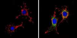
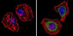
- Immunofluorescent analysis of Survivin (green) showing staining in the cytoplasm of C6 cells (right) compared to a negative control without primary antibody (left). Formalin-fixed cells were permeabilized with 0.1% Triton X-100 in TBS for 5-10 minutes and blocked with 3% BSA-PBS for 30 minutes at room temperature. Cells were probed with a Survivin monoclonal antibody (Product # MA5-11680) in 3% BSA-PBS at a dilution of 1:100 and incubated overnight at 4 ºC in a humidified chamber. Cells were washed with PBST and incubated with a DyLight-conjugated secondary antibody in PBS at room temperature in the dark. F-actin (red) was stained with a fluorescent red phalloidin and nuclei (blue) were stained with Hoechst or DAPI. Images were taken at a magnification of 60x.
- Submitted by
- Invitrogen Antibodies (provider)
- Main image
- Experimental details
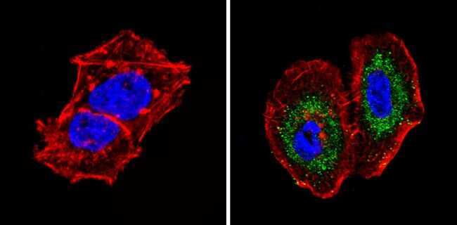
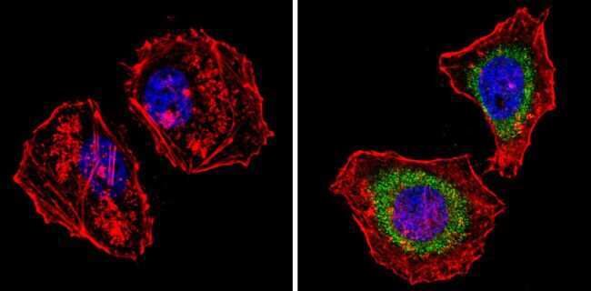
- Immunofluorescent analysis of Survivin (green) showing staining in the cytoplasm of Hela cells (right) compared to a negative control without primary antibody (left). Formalin-fixed cells were permeabilized with 0.1% Triton X-100 in TBS for 5-10 minutes and blocked with 3% BSA-PBS for 30 minutes at room temperature. Cells were probed with a Survivin monoclonal antibody (Product # MA5-11680) in 3% BSA-PBS at a dilution of 1:100 and incubated overnight at 4 ºC in a humidified chamber. Cells were washed with PBST and incubated with a DyLight-conjugated secondary antibody in PBS at room temperature in the dark. F-actin (red) was stained with a fluorescent red phalloidin and nuclei (blue) were stained with Hoechst or DAPI. Images were taken at a magnification of 60x.
Supportive validation
- Submitted by
- Invitrogen Antibodies (provider)
- Main image
- Experimental details
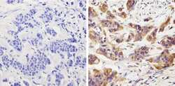
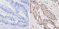
- Immunohistochemistry analysis of Survivin showing positive staining in the cytoplasm and nucleus of paraffin-treated Human bladder carcinoma (right) compared with a negative control in the absence of primary antibody (left). To expose target proteins, antigen retrieval method was performed using 10mM sodium citrate (pH 6.0) microwaved for 8-15 min. Following antigen retrieval, tissues were blocked in 3% H2O2-methanol for 15 min at room temperature, washed with ddH2O and PBS, and then probed with a Survivin monoclonal antibody (Product # MA5-11680) diluted by 3% BSA-PBS at a dilution of 1:50 overnight at 4°C in a humidified chamber. Tissues were washed extensively PBST and detection was performed using an HRP-conjugated secondary antibody followed by colorimetric detection using a DAB kit. Tissues were counterstained with hematoxylin and dehydrated with ethanol and xylene to prep for mounting.
- Submitted by
- Invitrogen Antibodies (provider)
- Main image
- Experimental details
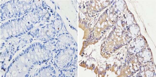
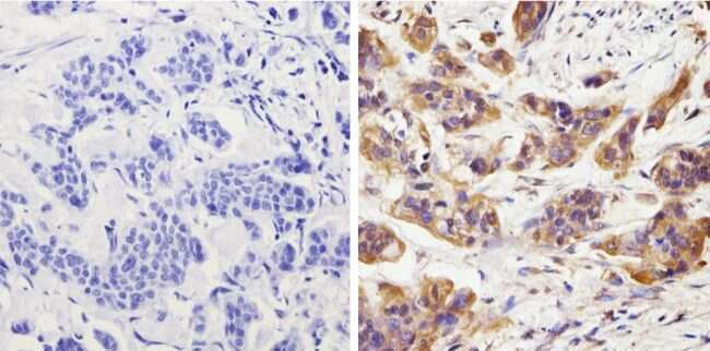
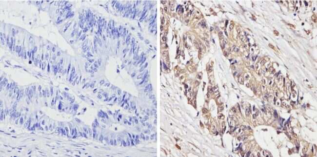
- Immunohistochemistry analysis of Survivin showing positive staining in the cytoplasm and nucleus of paraffin-treated Human colon carcinoma (right) compared with a negative control in the absence of primary antibody (left). To expose target proteins, antigen retrieval method was performed using 10mM sodium citrate (pH 6.0) microwaved for 8-15 min. Following antigen retrieval, tissues were blocked in 3% H2O2-methanol for 15 min at room temperature, washed with ddH2O and PBS, and then probed with a Survivin monoclonal antibody (Product # MA5-11680) diluted by 3% BSA-PBS at a dilution of 1:50 overnight at 4°C in a humidified chamber. Tissues were washed extensively PBST and detection was performed using an HRP-conjugated secondary antibody followed by colorimetric detection using a DAB kit. Tissues were counterstained with hematoxylin and dehydrated with ethanol and xylene to prep for mounting.
- Submitted by
- Invitrogen Antibodies (provider)
- Main image
- Experimental details
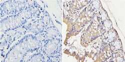
- Immunohistochemistry analysis of Survivin showing positive staining in the cytoplasm and nucleus of paraffin-treated Rat colon tissue (right) compared with a negative control in the absence of primary antibody (left). To expose target proteins, antigen retrieval method was performed using 10mM sodium citrate (pH 6.0) microwaved for 8-15 min. Following antigen retrieval, tissues were blocked in 3% H2O2-methanol for 15 min at room temperature, washed with ddH2O and PBS, and then probed with a Survivin monoclonal antibody (Product # MA5-11680) diluted by 3% BSA-PBS at a dilution of 1:50 overnight at 4°C in a humidified chamber. Tissues were washed extensively PBST and detection was performed using an HRP-conjugated secondary antibody followed by colorimetric detection using a DAB kit. Tissues were counterstained with hematoxylin and dehydrated with ethanol and xylene to prep for mounting.