 Explore
 Validate
 Learn
Learn Western blot
 Immunocytochemistry
Antibody data
- Antibody Data
- Antigen structure
- References [8]
- Comments [0]
- Validations
- Western blot [1]
Submit
Validation data
Reference
Comment
Report error
- Product number
- MAB6215 - Provider product page

- Provider
- Novus Biologicals
- Product name
- Mouse Monoclonal Caspase-1 Antibody
- Antibody type
- Monoclonal
- Description
- Protein A or G purified from hybridoma culture supernatant. Detects human Caspase-1 in direct ELISAs and Western blots.
- Reactivity
- Human
- Host
- Mouse
- Conjugate
- Unconjugated
- Isotype
- IgG
- Vial size
- 100 ug
- Concentration
- LYOPH
- Storage
- Use a manual defrost freezer and avoid repeated freeze-thaw cycles. 12 months from date of receipt, -20 to -70 degreesC as supplied. 1 month, 2 to 8 degreesC under sterile conditions after reconstitution. 6 months, -20 to -70 degreesC under sterile conditions after reconstitution.
Submitted references IL-1β activation in response to Staphylococcus aureus lung infection requires inflammasome-dependent and independent mechanisms.
The Vasoreparative Function of Myeloid Angiogenic Cells Is Impaired in Diabetes Through the Induction of IL1β.
Targeting the deubiquitinase STAMBP inhibits NALP7 inflammasome activity.
AIM2 Inflammasome Is Critical for Influenza-Induced Lung Injury and Mortality.
HMGB1 Induces an Inflammatory Response in the Chorioamniotic Membranes That Is Partially Mediated by the Inflammasome.
Monocyte Caspase-1 Is Released in a Stable, Active High Molecular Weight Complex Distinct from the Unstable Cell Lysate-Activated Caspase-1.
Age-related increases in amyloid beta and membrane attack complex: evidence of inflammasome activation in the rodent eye.
Propionibacterium acnes Induces IL-1β secretion via the NLRP3 inflammasome in human monocytes.
Pires S, Parker D
European journal of immunology 2018 Oct;48(10):1707-1716
European journal of immunology 2018 Oct;48(10):1707-1716
The Vasoreparative Function of Myeloid Angiogenic Cells Is Impaired in Diabetes Through the Induction of IL1β.
Chambers SEJ, O'Neill CL, Guduric-Fuchs J, McLoughlin KJ, Liew A, Egan AM, O'Brien T, Stitt AW, Medina RJ
Stem cells (Dayton, Ohio) 2018 Jun;36(6):834-843
Stem cells (Dayton, Ohio) 2018 Jun;36(6):834-843
Targeting the deubiquitinase STAMBP inhibits NALP7 inflammasome activity.
Bednash JS, Weathington N, Londino J, Rojas M, Gulick DL, Fort R, Han S, McKelvey AC, Chen BB, Mallampalli RK
Nature communications 2017 May 11;8:15203
Nature communications 2017 May 11;8:15203
AIM2 Inflammasome Is Critical for Influenza-Induced Lung Injury and Mortality.
Zhang H, Luo J, Alcorn JF, Chen K, Fan S, Pilewski J, Liu A, Chen W, Kolls JK, Wang J
Journal of immunology (Baltimore, Md. : 1950) 2017 Jun 1;198(11):4383-4393
Journal of immunology (Baltimore, Md. : 1950) 2017 Jun 1;198(11):4383-4393
HMGB1 Induces an Inflammatory Response in the Chorioamniotic Membranes That Is Partially Mediated by the Inflammasome.
Plazyo O, Romero R, Unkel R, Balancio A, Mial TN, Xu Y, Dong Z, Hassan SS, Gomez-Lopez N
Biology of reproduction 2016 Dec;95(6):130
Biology of reproduction 2016 Dec;95(6):130
Monocyte Caspase-1 Is Released in a Stable, Active High Molecular Weight Complex Distinct from the Unstable Cell Lysate-Activated Caspase-1.
Shamaa OR, Mitra S, Gavrilin MA, Wewers MD
PloS one 2015;10(11):e0142203
PloS one 2015;10(11):e0142203
Age-related increases in amyloid beta and membrane attack complex: evidence of inflammasome activation in the rodent eye.
Zhao T, Gao J, Van J, To E, Wang A, Cao S, Cui JZ, Guo JP, Lee M, McGeer PL, Matsubara JA
Journal of neuroinflammation 2015 Jun 24;12:121
Journal of neuroinflammation 2015 Jun 24;12:121
Propionibacterium acnes Induces IL-1β secretion via the NLRP3 inflammasome in human monocytes.
Qin M, Pirouz A, Kim MH, Krutzik SR, Garbán HJ, Kim J
The Journal of investigative dermatology 2014 Feb;134(2):381-388
The Journal of investigative dermatology 2014 Feb;134(2):381-388
No comments: Submit comment
Supportive validation
- Submitted by
- Novus Biologicals (provider)
- Main image
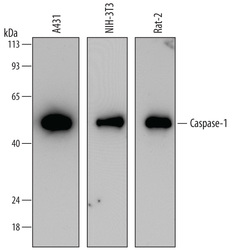
- Experimental details
- Detection of Human, Mouse, and Rat Caspase-1 by Western Blot. Western blot shows lysates of A431 human epithelial carcinoma cell line, NIH-3T3 mouse embryonic fibroblast cell line, and Rat-2 rat embryonic fibroblast cell line. PVDF membrane was probed with 0.1 µg/mL of Mouse Anti-Human Caspase-1 Monoclonal Antibody (Catalog # MAB6215) followed by HRP-conjugated Anti-Mouse IgG Secondary Antibody (Catalog # HAF007). A specific band was detected for Caspase-1 at approximately 45 kDa (as indicated). This experiment was conducted under reducing conditions and using Immunoblot Buffer Group 2.
