Explore
 Validate
 Learn
Learn Western blot
 Immunocytochemistry
Antibody data
- Antibody Data
- Antigen structure
- References [2]
- Comments [0]
- Validations
- Immunocytochemistry [2]
- Other assay [4]
Submit
Validation data
Reference
Comment
Report error
- Product number
- PA5-87536 - Provider product page

- Provider
- Invitrogen Antibodies
- Product name
- Caspase 1 Polyclonal Antibody
- Antibody type
- Polyclonal
- Antigen
- Synthetic peptide
- Description
- Positive Samples: THP-1, Mouse lung, Rat spleen; Cellular Location: Cytoplasm Immunogen sequence: CRGDSPGVVW FK
- Reactivity
- Human, Mouse
- Host
- Rabbit
- Isotype
- IgG
- Vial size
- 100 µL
- Concentration
- 2.93 mg/mL
- Storage
- -20° C, Avoid Freeze/Thaw Cycles
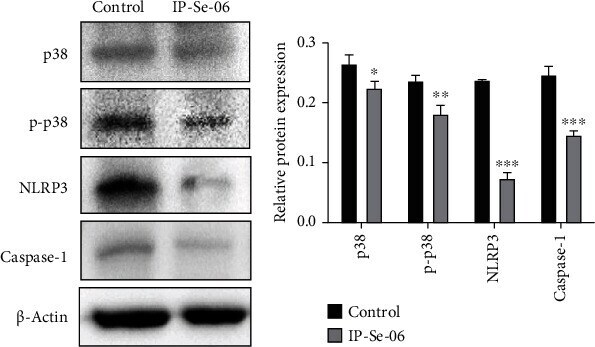
Submitted references IP-Se-06, a Selenylated Imidazo[1,2-a]pyridine, Modulates Intracellular Redox State and Causes Akt/mTOR/HIF-1α and MAPK Signaling Inhibition, Promoting Antiproliferative Effect and Apoptosis in Glioblastoma Cells.
Modulation of Gut Microbiota Combined with Upregulation of Intestinal Tight Junction Explains Anti-Inflammatory Effect of Corylin on Colitis-Associated Cancer in Mice.
Dos Santos DC, Rafique J, Saba S, Grinevicius VMAS, Filho DW, Zamoner A, Braga AL, Pedrosa RC, Ourique F
Oxidative medicine and cellular longevity 2022;2022:3710449
Oxidative medicine and cellular longevity 2022;2022:3710449
Modulation of Gut Microbiota Combined with Upregulation of Intestinal Tight Junction Explains Anti-Inflammatory Effect of Corylin on Colitis-Associated Cancer in Mice.
Chang ZY, Liu HM, Leu YL, Hsu CH, Lee TY
International journal of molecular sciences 2022 Feb 28;23(5)
International journal of molecular sciences 2022 Feb 28;23(5)
No comments: Submit comment
Supportive validation
- Submitted by
- Invitrogen Antibodies (provider)
- Main image
- Experimental details
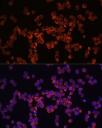
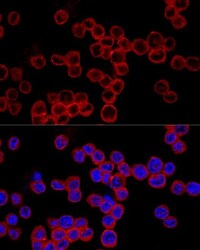
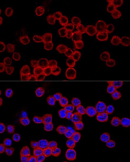
- Immunocytochemistry-Immunofluorescence analysis of Caspase 1 was performed in Raw264.7 cells using Caspase 1 Polyclonal Antibody (Product # PA5-87536) at a dilution of 1:100. Blue: DAPI for nuclear staining.
- Submitted by
- Invitrogen Antibodies (provider)
- Main image
- Experimental details
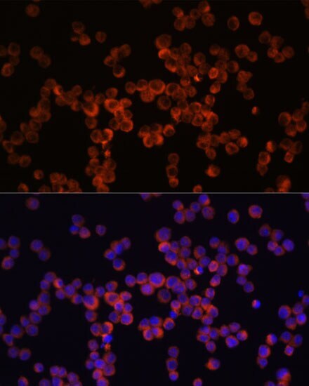
- Immunocytochemistry-Immunofluorescence analysis of Caspase 1 was performed in Raw264.7 cells using Caspase 1 Polyclonal Antibody (Product # PA5-87536) at a dilution of 1:200. Blue: DAPI for nuclear staining.
Supportive validation
- Submitted by
- Invitrogen Antibodies (provider)
- Main image
- Experimental details
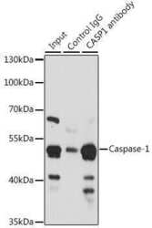
- Immunoprecipitation analysis of Caspase 1 was performed in 200 µg extracts of THP-1 cells using Caspase 1 Polyclonal Antibody (Product # PA5-87536). Western blot was performed from the immunoprecipitate using Caspase 1 Polyclonal Antibody.

- Submitted by
- Invitrogen Antibodies (provider)
- Main image

- Experimental details
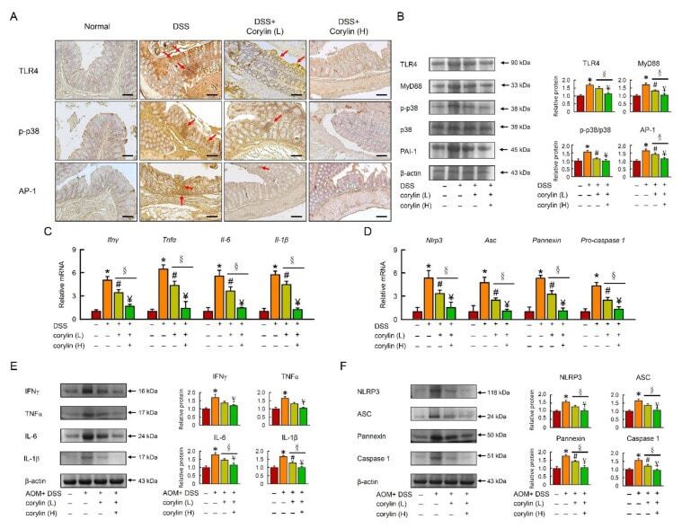
- Corylin improves the TLR4 signal pathway in DSS-induced colitis mice. ( A ) Expression analysis by immunohistochemical staining of TLR4, p-p38, and AP-1 in mice after the indicated treatment. Scale bar: 100 mum. Red arrows highlight positive staining. ( B ) Western blot analysis of TLR4, MyD88, p-p38, AP-1, and beta-actin (loading control) in colon homogenates. Right graph indicates quantification relative to beta-actin. ( C , D ) mRNA expression of Ifngamma , Tnf-alpha , Il-6 , Il-1beta , Nlrp3 , Asc , Pannexin , and Pro-caspase 1 determined by qRT-PCR. ( E , F ) Western blot analysis of IFNgamma, TNF-alpha, IL-6, IL-1beta, NLRP3, ASC, Pannexin, and Caspase 1, and beta-actin (loading control) in colon homogenates. Results represent mean +- SEM. * p < 0.05, Normal compared with DSS; # p < 0.05, DSS compared with DSS + Corylin (L); Y= p < 0.05, DSS compared with DSS + Corylin (H); SS p < 0.05, DSS + Corylin (L) compared with DSS + Corylin (H). DSS, dextran sodium sulfate; TLR4, Toll-like receptor 4; MyD88, myeloid differentiation primary response 88; AP-1, activator protein 1; Ifngamma , interferon gamma; Tnf-alpha , tumor necrosis factor-alpha; Il-6 , interleukin-6; Nlrp3 , NACHT, LRR, and PYD domains-containing protein 3.
- Submitted by
- Invitrogen Antibodies (provider)
- Main image
- Experimental details
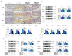
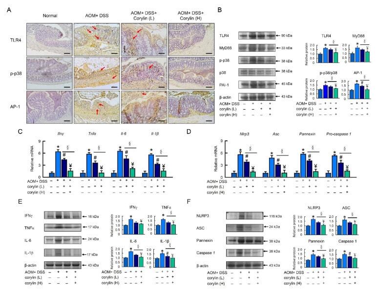
- Corylin improves the TLR4 signaling pathway in AOM/DSS-induced-colitis-associated colorectal cancer mice. ( A ) Expression analysis by immunohistochemical staining of TLR4, p-p38, and AP-1 in mice after the indicated treatment. Scale bar: 100 mum. ( B ) Western blot analysis of TLR4, MyD88, p-p38, AP-1, and beta-actin (loading control) in colon. ( C , D ) mRNA expression of Ifngamma , Tnf-alpha , Il-6 , Il-1beta , Nlrp3 , Asc , Pannexin , and Pro-caspase 1 as determined by qRT-PCR. ( E , F ) Western blot analysis of IFNgamma, TNF-alpha, IL-6, IL-1beta, NLRP3, ASC, Pannexin, and Caspase 1, and beta-actin (loading control) in colon homogenates. Results represent mean +- SEM. * p < 0.05, Normal compared with AOM/DSS; # p < 0.05, AOM/DSS compared with AOM/DSS + Corylin (L); Y= p < 0.05, AOM/DSS compared with AOM/DSS + Corylin (H); SS p < 0.05, DSS + Corylin (L) compared with DSS + Corylin (H). AOM, azoxymethane; DSS, dextran sodium sulfate; TLR4, Toll-like receptor 4; MyD88, myeloid differentiation primary response 88; AP-1, activator protein 1; Ifngamma , interferon gamma; Tnf-alpha , tumor necrosis factor-alpha; Il-6 , interleukin-6; Nlrp3 , NACHT, LRR, and PYD domains-containing protein 3.
- Submitted by
- Invitrogen Antibodies (provider)
- Main image
- Experimental details
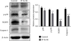
- Treatment with IP-Se-06 (1 mu M) at 48 h downregulated p38 mitogen-activated protein kinase and inflammasome complex protein in A172 glioblastoma cells. (a) Immunoblotting analysis detected the downregulation of p38, p-p38, NLRP3 and caspase-1 protein levels. beta -Actin was used as a loading control. (b) Densitometric analysis results of p38, p-p38, NLRP3 and caspase-1 were quantified with ImageJ. The data were considered as statistically significant at * p < 0.05, ** p < 0.01, and *** p < 0.001 compared to the control group (nontreated cells).