Explore
 Validate
 Learn
Learn Western blot
 Immunohistochemistry
Antibody data
- Antibody Data
- Antigen structure
- References [1]
- Comments [0]
- Validations
- Immunohistochemistry [6]
- Other assay [2]
Submit
Validation data
Reference
Comment
Report error
- Product number
- MA5-16215 - Provider product page

- Provider
- Invitrogen Antibodies
- Product name
- Caspase 1 Monoclonal Antibody (14F468)
- Antibody type
- Monoclonal
- Antigen
- Synthetic peptide
- Description
- Suggested positive control: 3T3 whole cell lysate, antigen standard for CASP1 (transient overexpression lysate), HeLa or NIH-3T3, Hela whole cell lysate. Immunogen's sequence similarity with other species: Porcine/Pig (85%), Equine/Horse (80%), Canine (70%). Staining of formalin-fixed tissues is enhanced by boiling tissue sections in 10 mM sodium citrate buffer, pH 6.0 for 10-20 min followed by cooling at RT for 20 min.
- Reactivity
- Human, Mouse, Rat
- Host
- Mouse
- Isotype
- IgG
- Antibody clone number
- 14F468
- Vial size
- 100 μg
- Concentration
- 1.0 mg/mL
- Storage
- Store at 4°C short term. For long term storage, store at -20°C, avoiding freeze/thaw cycles.
Submitted references Gasdermin D: Evidence of pyroptosis in spontaneous preterm labor with sterile intra-amniotic inflammation or intra-amniotic infection.
Gomez-Lopez N, Romero R, Tarca AL, Miller D, Panaitescu B, Schwenkel G, Gudicha DW, Hassan SS, Pacora P, Jung E, Hsu CD
American journal of reproductive immunology (New York, N.Y. : 1989) 2019 Dec;82(6):e13184
American journal of reproductive immunology (New York, N.Y. : 1989) 2019 Dec;82(6):e13184
No comments: Submit comment
Supportive validation
- Submitted by
- Invitrogen Antibodies (provider)
- Main image

- Experimental details
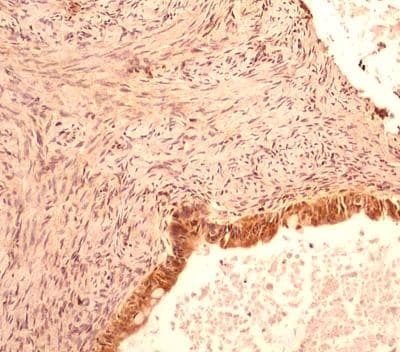
- Immunohistochemical analysis of Caspase 1 in Adenocarcinoma of the rectum. Samples were incubated in Caspase 1 monoclonal antibody (Product # MA5-16215) using a dilution of 5 µg/mL followed by a peroxidase-conjugate and DAB chromogen. Staining of formalin-fixed tissues is enhanced by boiling tissue sections in 10 mM sodium citrate buffer, pH 6.0 for 10-20 min followed by cooling at RT for 20 min.
- Submitted by
- Invitrogen Antibodies (provider)
- Main image

- Experimental details
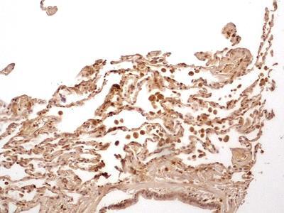
- Immunohistochemical analysis of Caspase 1 in Normal lung from human. Samples were incubated in Caspase 1 monoclonal antibody (Product # MA5-16215) using a dilution of 5 µg/mL. In this representative lung section, different type of cells including pseudostratified columnar epithelium of bronchiole and the simple squamous epithelium of alveoli may be seen to develop immunoreactivity for Caspase 1. [10X Magnification].
- Submitted by
- Invitrogen Antibodies (provider)
- Main image

- Experimental details
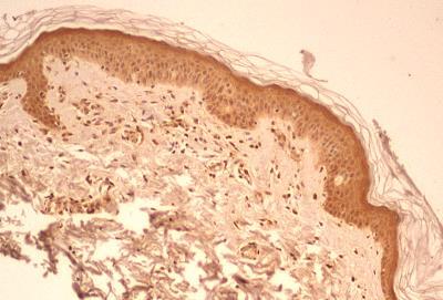
- Immunohistochemical analysis of Caspase 1 in Normal skin from human. Samples were incubated in Caspase 1 monoclonal antibody (Product # MA5-16215) using a dilution of 5 µg/mL. Strong cytoplasmic/nuclear staining developed in all the epidermal cells, blood vessels and some cells of the dermal connective tissues layer. [10X Magnification].
- Submitted by
- Invitrogen Antibodies (provider)
- Main image
- Experimental details
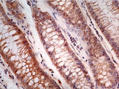
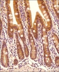
- Immunohistochemical analysis of Caspase 1 in Tissue section of human intestine. Samples were incubated in Caspase 1 monoclonal antibody (Product # MA5-16215) using a dilution of 5 µg/mL. The primary antibody binding to Caspase 1 in cells was detected using HRP conjugated anti-Mouse secondary antibody with DAB reagent, and the sections were further counterstained with hematoxylin for labeling cellular nuclei. This Caspase 1 antibody generated a diffused but specific cytoplasmic staining in columnar epithelia cells of villi, and a few cells depicted nuclear staining also. Only a subset of connective tissue cells in lamina propria depicted positivity (cytoplasmic) for this protein.

- Submitted by
- Invitrogen Antibodies (provider)
- Main image

- Experimental details
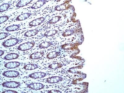
- Immunohistochemical analysis of Caspase 1 in a section of normal human colon. Samples were incubated in Caspase 1 monoclonal antibody (Product # MA5-16215) using a dilution of 5 µg/mL. Distinct cytoplasmic staining along with some nuclear positivity was observed in crypts/mucosa, and staining was found to be more intense in the absorptive columnar epithelial cells. [10X Magnification].
- Submitted by
- Invitrogen Antibodies (provider)
- Main image
- Experimental details
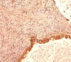
- Immunohistochemical analysis of Caspase 1 in a section of human ovarian cancer. Samples were incubated in Caspase 1 monoclonal antibody (Product # MA5-16215) using a dilution of 5 µg/mL.
Supportive validation
- Submitted by
- Invitrogen Antibodies (provider)
- Main image
- Experimental details
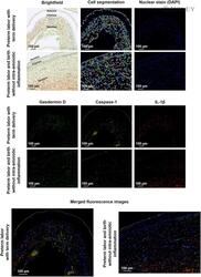
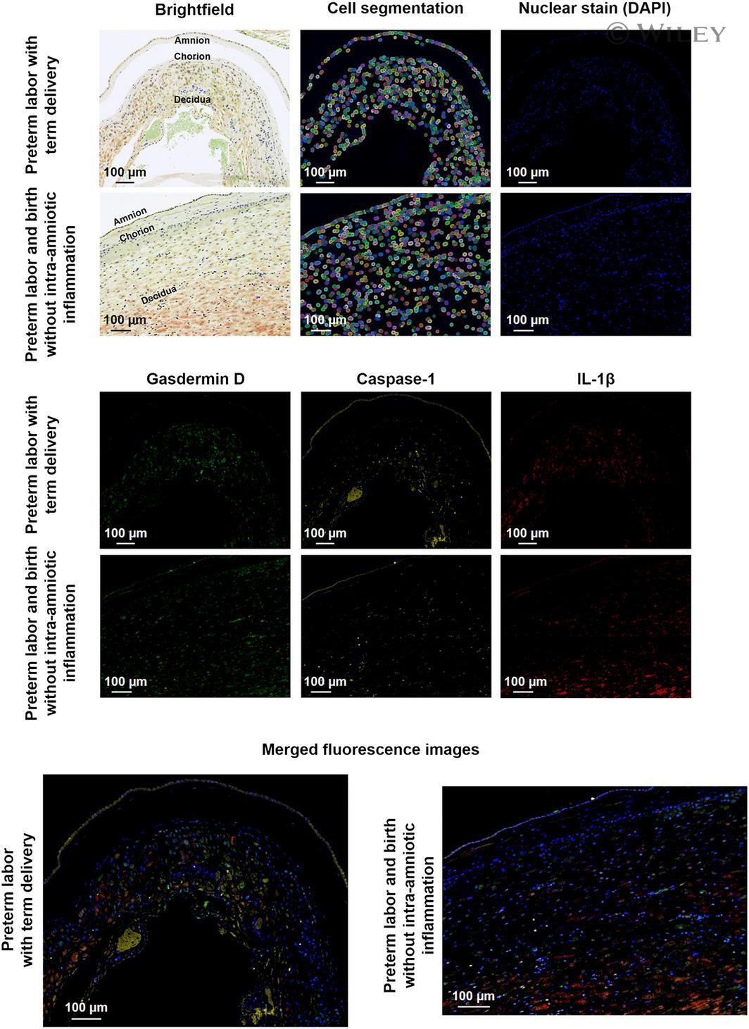
- Gasdermin D expression in the chorioamniotic membranes of women who underwent spontaneous preterm labor without intra-amniotic inflammation. Representative multiplex immunofluorescence images showing the brightfield view, cell segmentation map, nuclear staining (4',6-diamidino-2-phenylindole, DAPI, blue), and protein expression of gasdermin D (green), caspase-1 (yellow), and IL-1beta (red) in the chorioamniotic membranes from women who underwent preterm labor and delivered at term (upper rows, left merged image) or preterm (bottom rows, right merged image) without intra-amniotic inflammation. Merged images show the co-localization of gasdermin D, caspase-1, and IL-1beta expression. Images taken at 200x magnification. Scale bars = 100 um
- Submitted by
- Invitrogen Antibodies (provider)
- Main image
- Experimental details
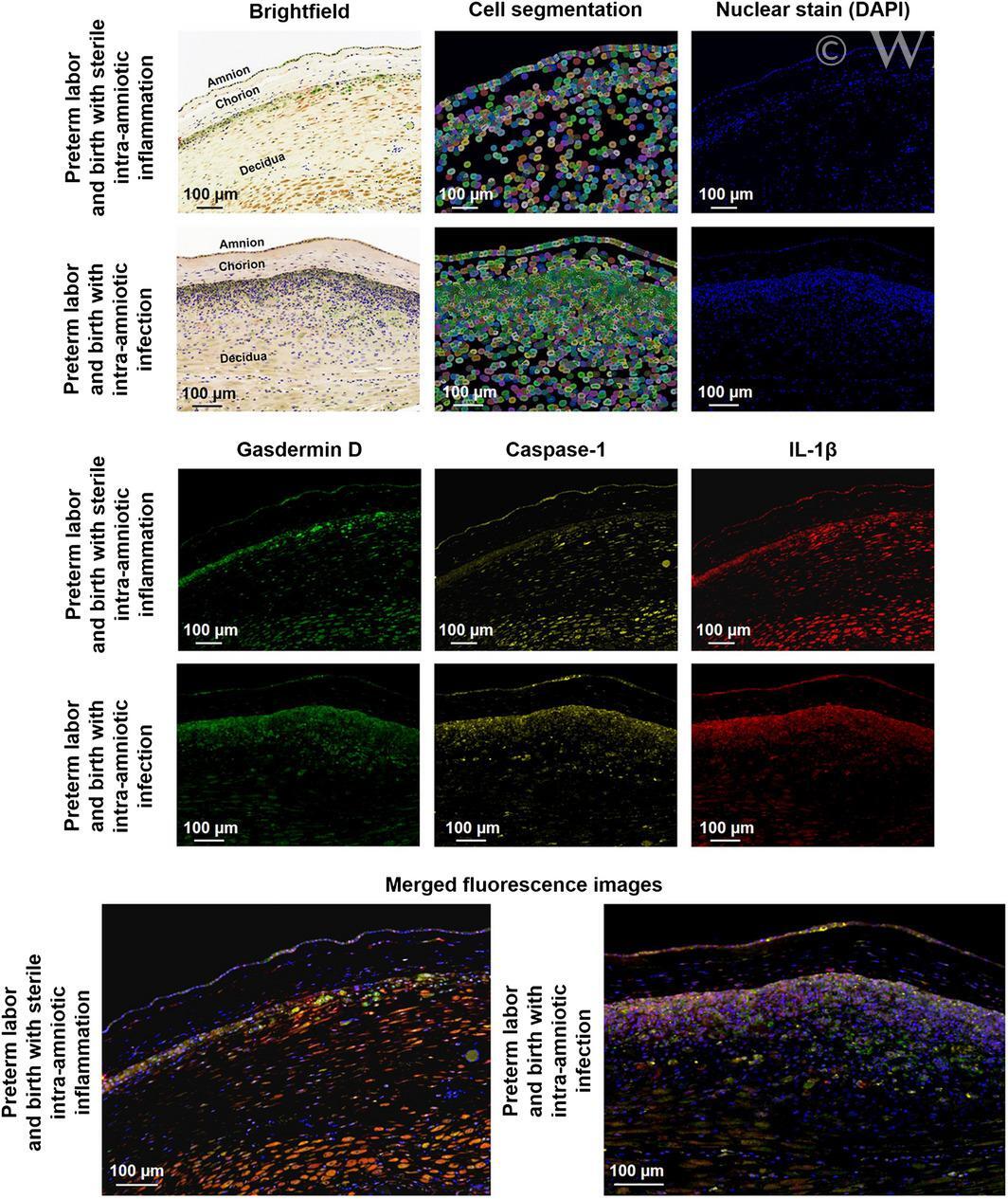
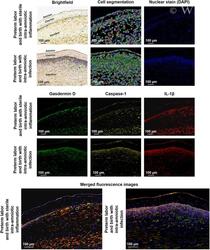
- Gasdermin D expression in the chorioamniotic membranes of women with spontaneous preterm labor and sterile intra-amniotic inflammation or intra-amniotic infection. Representative multiplex immunofluorescence images showing the brightfield view, cell segmentation map, nuclear staining (4',6-diamidino-2-phenylindole, DAPI, blue), and protein expression of gasdermin D (green), caspase-1 (yellow), and IL-1beta (red) in the chorioamniotic membranes from women who underwent preterm labor and birth with sterile intra-amniotic inflammation (upper rows, left merged image) or intra-amniotic infection (bottom rows, right merged image). Merged images show the co-localization of gasdermin D, caspase-1, and IL-1beta expression. Images taken at 200x magnification. Scale bars = 100 um