 Explore
 Validate
 Learn
Learn Western blot
 Immunocytochemistry
Antibody data
- Antibody Data
- Antigen structure
- References [1]
- Comments [0]
- Validations
- Immunocytochemistry [3]
- Immunohistochemistry [1]
- Other assay [1]
Submit
Validation data
Reference
Comment
Report error
- Product number
- PA5-20113 - Provider product page

- Provider
- Invitrogen Antibodies
- Product name
- Caspase 1 Polyclonal Antibody
- Antibody type
- Polyclonal
- Antigen
- Synthetic peptide
- Description
- A suggested positive control is Hela cell lysate. PA5-20113 can be used with blocking peptide PEP-0231.
- Reactivity
- Human
- Host
- Rabbit
- Isotype
- IgG
- Vial size
- 100 μg
- Concentration
- 1 mg/mL
- Storage
- Maintain refrigerated at 2-8°C for up to 3 months. For long term storage store at -20°C
Submitted references Myocardial hypothermia increases autophagic flux, mitochondrial mass and myocardial function after ischemia-reperfusion injury.
Marek-Iannucci S, Thomas A, Hou J, Crupi A, Sin J, Taylor DJ, Czer LS, Esmailian F, Mentzer RM Jr, Andres AM, Gottlieb RA
Scientific reports 2019 Jul 10;9(1):10001
Scientific reports 2019 Jul 10;9(1):10001
No comments: Submit comment
Supportive validation
- Submitted by
- Invitrogen Antibodies (provider)
- Main image
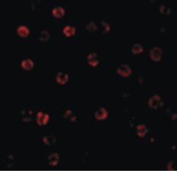
- Experimental details
- Immunofluorescent analysis of HeLa cells using a Caspase-1 polyclonal antibody (Product # PA5-20113) at a 20 µg/mL dilution.
- Submitted by
- Invitrogen Antibodies (provider)
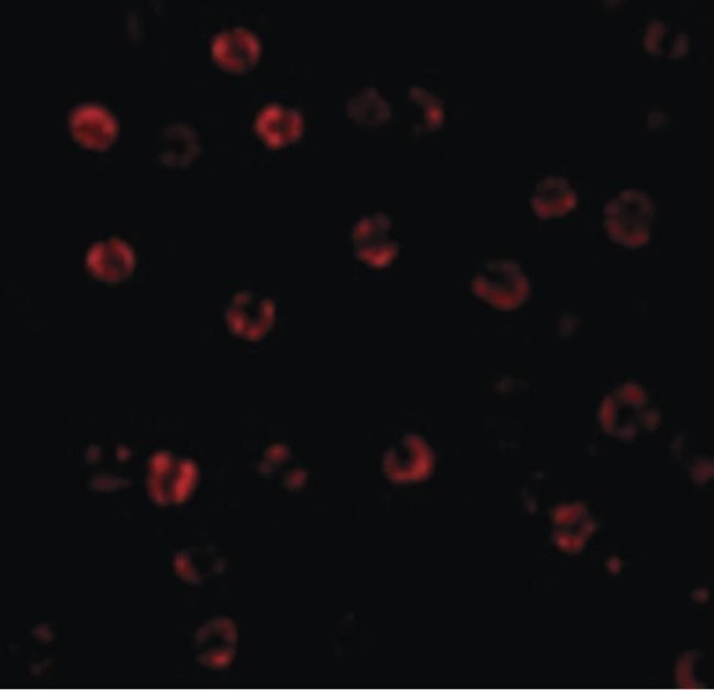
- Main image
- Experimental details
- Immunofluorescence of Caspase-1 in Hela cells with Caspase 1 Polyclonal Antibody (Product # PA5-20113) at 20 µg/mL.

- Submitted by
- Invitrogen Antibodies (provider)
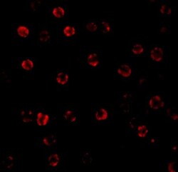
- Main image

- Experimental details
- Immunofluorescence of Caspase-1 in Hela cells with Caspase 1 Polyclonal Antibody (Product # PA5-20113) at 20 µg/mL.

Supportive validation
- Submitted by
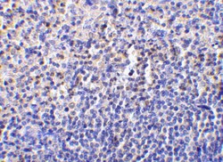
- Invitrogen Antibodies (provider)
- Main image
- Experimental details
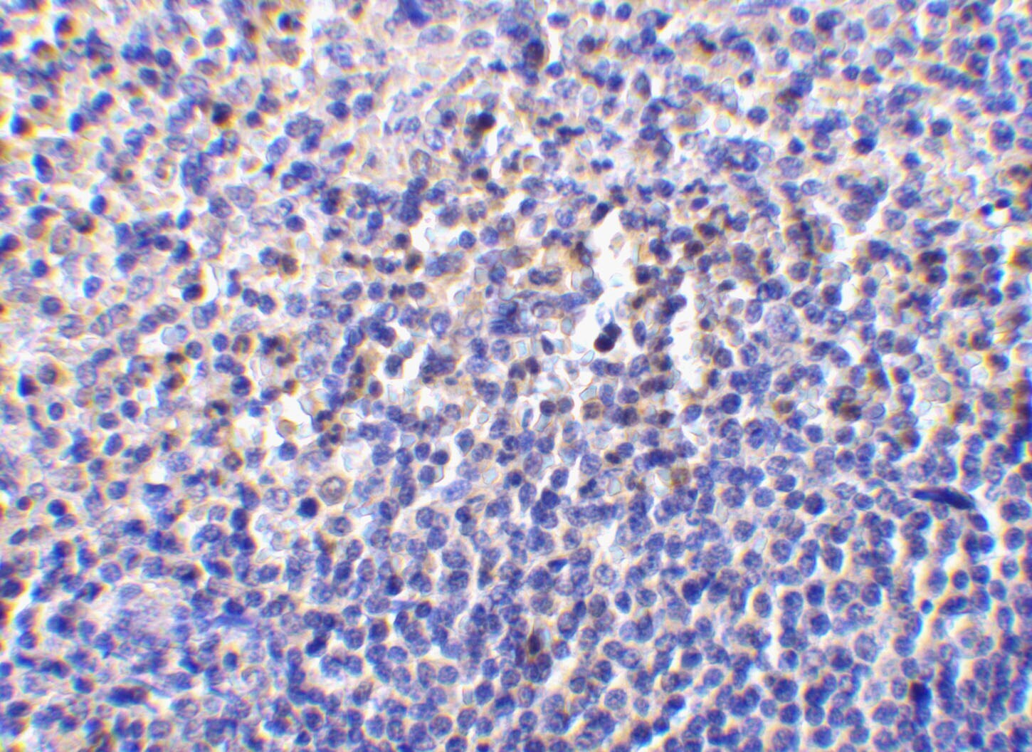
- Immunohistochemistry of caspase-1 in human spleen tissue with Caspase 1 Polyclonal Antibody (Product # PA5-20113) at 10 µg/mL.
Supportive validation
- Submitted by
- Invitrogen Antibodies (provider)
- Main image
- Experimental details
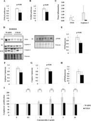
- Figure 8 Myocardial hypothermia decreases local and systemic inflammation. Myocardial border zone tissue of female farm pigs (n = 3 per group) was harvested one week post MI. ( A , B ) Quantification of TNFalpha and TGFbeta mRNA expression respectively. ( C ) Quantification of local tissue inflammation via iNOS and Arginase-1 staining (representative images: Supplemental Fig. 2 ). ( D ) WB analysis with protein quantification ( E - G ) of iNOS, Arginase-1 and Caspase-1 respectively in whole lysate and ( H ) pNFkappaB in the nuclear fraction. ( I ) Quantification of lymphocyte proliferation with Concanavalin-A treatment on fresh splenic tissue, harvested from the same pigs. The data represents the mean +- SEM with *p < 0.05, **p < 0.01, comparing cold (black) vs. warm (white). We normalized each protein to the entire corresponding lane on the ponceau-stained membrane, representing the total protein amount. Due to space limitation, the blots are cropped from various gels, please see the supplemental data for full-length gels/blots.
