Explore
 Validate
 Learn
Learn Western blot
Antibody data
- Antibody Data
- Antigen structure
- References [3]
- Comments [0]
- Validations
- Western blot [4]
- Immunocytochemistry [1]
Submit
Validation data
Reference
Comment
Report error
- Product number
- AF6215 - Provider product page

- Provider
- R&D Systems
- Product name
- Human Caspase-1 Antibody
- Antibody type
- Polyclonal
- Description
- Antigen Affinity-purified. Detects human Caspase-1 in Western blots.
- Reactivity
- Human
- Host
- Goat
- Conjugate
- Unconjugated
- Antigen sequence
P29466- Isotype
- IgG
- Vial size
- 100 ug
- Concentration
- LYOPH
- Storage
- Use a manual defrost freezer and avoid repeated freeze-thaw cycles. 12 months from date of receipt, -20 to -70 °C as supplied. 1 month, 2 to 8 °C under sterile conditions after reconstitution. 6 months, -20 to -70 °C under sterile conditions after reconstitution.
Submitted references Human metapneumovirus activates NOD-like receptor protein 3 inflammasome via its small hydrophobic protein which plays a detrimental role during infection in mice.
Cutting Edge: Inflammasome Activation in Primary Human Macrophages Is Dependent on Flagellin.
Staphylococcus aureus activates the NLRP3 inflammasome in human and rat conjunctival goblet cells.
Lê VB, Dubois J, Couture C, Cavanagh MH, Uyar O, Pizzorno A, Rosa-Calatrava M, Hamelin MÈ, Boivin G
PLoS pathogens 2019 Apr;15(4):e1007689
PLoS pathogens 2019 Apr;15(4):e1007689
Cutting Edge: Inflammasome Activation in Primary Human Macrophages Is Dependent on Flagellin.
Kortmann J, Brubaker SW, Monack DM
Journal of immunology (Baltimore, Md. : 1950) 2015 Aug 1;195(3):815-9
Journal of immunology (Baltimore, Md. : 1950) 2015 Aug 1;195(3):815-9
Staphylococcus aureus activates the NLRP3 inflammasome in human and rat conjunctival goblet cells.
McGilligan VE, Gregory-Ksander MS, Li D, Moore JE, Hodges RR, Gilmore MS, Moore TC, Dartt DA
PloS one 2013;8(9):e74010
PloS one 2013;8(9):e74010
No comments: Submit comment
Supportive validation
- Submitted by
- R&D Systems (provider)
- Main image

- Experimental details
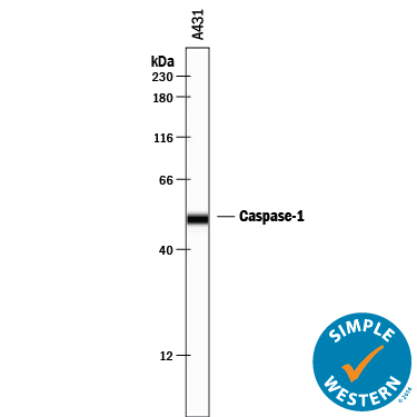
- Detection of Human Caspase-1 by Simple WesternTM. Simple Western lane view shows lysates of A431 human epithelial carcinoma cell line, loaded at 0.2 mg/mL. A specific band was detected for Caspase-1 at approximately 51 kDa (as indicated) using 12.5 µg/mL of Goat Anti-Human Caspase-1 Antigen Affinity-purified Polyclonal Antibody (Catalog # AF6215) followed by 1:50 dilution of HRP-conjugated Anti-Goat IgG Secondary Antibody (Catalog # HAF109). This experiment was conducted under reducing conditions and using the 12-230 kDa separation system.
- Submitted by
- R&D Systems (provider)
- Main image

- Experimental details
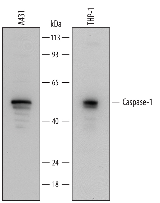
- Detection of Human Caspase-1 by Western Blot. Western blot shows lysates of A431 human epithelial carcinoma cell line and THP-1 human acute monocytic leukemia cell line. PVDF Membrane was probed with 0.25 µg/mL of Goat Anti-Human Caspase-1 Antigen Affinity-purified Polyclonal Antibody (Catalog # AF6215) followed by HRP-conjugated Anti-Goat IgG Secondary Antibody (Catalog # HAF109). A specific band was detected for Caspase-1 at approximately 50 kDa (as indicated). This experiment was conducted under reducing conditions and using Immunoblot Buffer Group 2.
- Submitted by
- R&D Systems (provider)
- Main image
- Experimental details
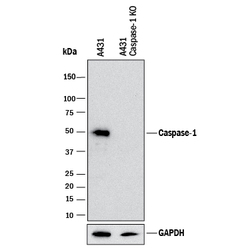
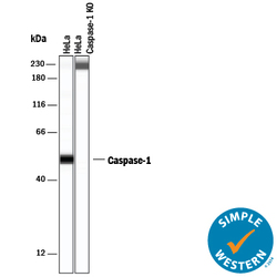
- Western Blot Shows Human Caspase-1 Specificity by Using Knockout Cell Line. Western blot shows lysates of A431 human epithelial carcinoma parental cell line and Caspase-1 knockout A431 cell line (KO). PVDF membrane was probed with 0.5 µg/mL of Goat Anti-Human Caspase-1 Antigen Affinity-purified Polyclonal Antibody (Catalog # AF6215) followed by HRP-conjugated Anti-Goat IgG Secondary Antibody (Catalog # HAF017). A specific band was detected for Caspase-1 at approximately 50 kDa (as indicated) in the parental A431 cell line, but is not detectable in knockout A431 cell line. GAPDH (Catalog # AF5718) is shown as a loading control. This experiment was conducted under reducing conditions and using Immunoblot Buffer Group 1.

- Submitted by
- R&D Systems (provider)
- Main image
- Experimental details
- Detection of Human Caspase-1 by Simple WesternTM. Simple Western lane view shows lysates of A431 human epithelial carcinoma cell line, loaded at 0.2 mg/mL. A specific band was detected for Caspase-1 at approximately 51 kDa (as indicated) using 12.5 µg/mL of Goat Anti-Human Caspase-1 Antigen Affinity-purified Polyclonal Antibody (Catalog # AF6215) followed by 1:50 dilution of HRP-conjugated Anti-Goat IgG Secondary Antibody (Catalog # HAF109). This experiment was conducted under reducing conditions and using the 12-230 kDa separation system.

Supportive validation
- Submitted by
- R&D Systems (provider)
- Main image

- Experimental details
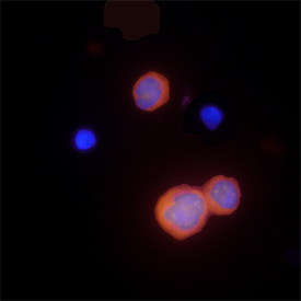
- Caspase-1 in THP-1 Human Cell Line. Caspase-1 was detected in immersion fixed THP-1 human acute monocytic leukemia cell line using Goat Anti-Human Caspase-1 Antigen Affinity-purified Polyclonal Antibody (Catalog # AF6215) at 15 µg/mL for 3 hours at room temperature. Cells were stained using the NorthernLights™ 557-conjugated Anti-Goat IgG Secondary Antibody (red; Catalog # NL001) and counterstained with DAPI (blue). Specific staining was localized to cytoplasm. View our protocol for Fluorescent ICC Staining of Non-adherent Cells.