 Explore
 Validate
 Learn
Learn Western blot
Antibody data
- Antibody Data
- Antigen structure
- References [1]
- Comments [0]
- Validations
- Western blot [3]
- Other assay [3]
Submit
Validation data
Reference
Comment
Report error
- Product number
- PA5-13799 - Provider product page

- Provider
- Invitrogen Antibodies
- Product name
- PRKAR2B Polyclonal Antibody
- Antibody type
- Polyclonal
- Antigen
- Synthetic peptide
- Reactivity
- Human, Mouse
- Host
- Rabbit
- Isotype
- IgG
- Vial size
- 400 µL
- Concentration
- 2 mg/mL
- Storage
- Store at 4°C short term. For long term storage, store at -20°C, avoiding freeze/thaw cycles.
Submitted references PRKAR2B plays an oncogenic role in the castration-resistant prostate cancer.
Sha J, Xue W, Dong B, Pan J, Wu X, Li D, Liu D, Huang Y
Oncotarget 2017 Jan 24;8(4):6114-6129
Oncotarget 2017 Jan 24;8(4):6114-6129
No comments: Submit comment
Supportive validation
- Submitted by
- Invitrogen Antibodies (provider)
- Main image
- Experimental details
- Western blot analysis using a PKA 2 beta polyclonal antibody (Product # PA5-13799) in mouse brain tissue lysate. Secondary HRP-anti-rabbit was used for signal visualization with chemiluminescence.

- Submitted by
- Invitrogen Antibodies (provider)
- Main image

- Experimental details
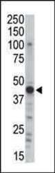
- Western blot analysis of PRKAR2B (arrow) using a PKA 2 beta polyclonal antibody (Product # PA5-13799) in 293 cell lysates (2 µg/lane) either nontransfected (Lane 1) or transiently transfected (Lane 2) with the PRKAR2B gene.

- Submitted by
- Invitrogen Antibodies (provider)
- Main image

- Experimental details
- Western blot analysis using a PKA 2 beta polyclonal antibody (Product # PA5-13799) in Ramos cell lysates (35 µg per lane).

Supportive validation
- Submitted by
- Invitrogen Antibodies (provider)
- Main image
- Experimental details
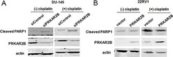
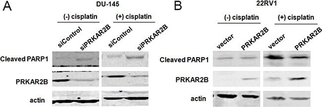
- Figure 5 PRKAR2B plays the role of anti-apoptosis in CRPC cells ( A ) DU145 cell was transfected with PRKAR2B siRNA for 48 hours, and then treated with DMSO (left panel) or 10 muM cisplatin (right panel) for 24 hours. The protein expression of PRKAR2B and cleaved PARP1 were examined through western blot. Actin was the internal loading control. ( B ) 22RV1 cells was transfected with PRKAR2B plasmid for 48 hours, and then treated with 10 muM cisplatin for 24 hours. The protein expression of PRKAR2B and cleaved PARP1 were examined through western blot. Actin was the internal loading control.
- Submitted by
- Invitrogen Antibodies (provider)
- Main image
- Experimental details
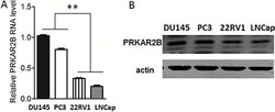
- Figure 2 Expression of PRKAR2B in prostate cancer cell lines ( A ) The mRNA expression level of PRKAR2B in DU145, PC3, 22RV1 and LNCap cells was detected by Real-Time qPCR. ( B ) The protein expression level of PRKAR2B in DU145, PC3, 22RV1 and LNCap cells was detected by western-blot, and beta-actin was the internal loading control (** P < 0.01).

- Submitted by
- Invitrogen Antibodies (provider)
- Main image

- Experimental details
- Figure 3 PRKAR2B promotes CRPC cell proliferation ( A ) The mRNA and protein expression level of PRKAR2B was examined by Real-Time qPCR (bar graph) and western blot, respectively, in DU145 and PC3 cells with or without PRKAR2B knockdown. ( B ) MTT assay shows cell viability of DU145 cells (upper panel) and PC3 cells (lower panel) withor without PRKAR2Bknockdown. OD value at 570nmwas tested at 0 h, 24 h, 48 h, 72 h and 96 h after siRNA transfection. siN: negative control siRNA;siPRKAR2B: PRKAR2B siRNA. ( C ) The mRNA and protein expression level of PRKAR2B was examined by Real-Time qPCR (bar graph) and western blot, respectively, in 22RV1 and LNCap cells after transfection of PRKAR2B plasmid for 48 hours. ( D ) MTT assay showed cell proliferation of 22RV1 cells(upper panel) and LNCap cells(lower panel) with or without PRKAR2B plasmid transfection. OD value at 570nmwas tested at 0 h, 24 h, 48 h, 72 h and 96 h after plasmid transfection (* P < 0.05; ** P < 0.01; *** P < 0.001).
