Explore
 Validate
 Learn
Learn Western blot
 Immunocytochemistry
Antibody data
- Antibody Data
- Antigen structure
- References [2]
- Comments [0]
- Validations
- Immunocytochemistry [1]
- Immunohistochemistry [2]
- Other assay [5]
Submit
Validation data
Reference
Comment
Report error
- Product number
- PA5-28266 - Provider product page

- Provider
- Invitrogen Antibodies
- Product name
- PRKAR2B Polyclonal Antibody
- Antibody type
- Polyclonal
- Antigen
- Recombinant full-length protein
- Description
- Recommended positive controls: HeLa, HepG2, mouse brain, rat brain. Predicted reactivity: Mouse (97%), Rat (96%), Xenopus laevis (84%), Bovine (99%). Store product as a concentrated solution. Centrifuge briefly prior to opening the vial.
- Reactivity
- Human, Mouse, Rat
- Host
- Rabbit
- Isotype
- IgG
- Vial size
- 100 μL
- Concentration
- 0.67 mg/mL
- Storage
- Store at 4°C short term. For long term storage, store at -20°C, avoiding freeze/thaw cycles.
Submitted references Melanoma-Secreted Amyloid Beta Suppresses Neuroinflammation and Promotes Brain Metastasis.
PRKAR2B-HIF-1α loop promotes aerobic glycolysis and tumour growth in prostate cancer.
Kleffman K, Levinson G, Rose IVL, Blumenberg LM, Shadaloey SAA, Dhabaria A, Wong E, Galán-Echevarría F, Karz A, Argibay D, Von Itter R, Floristán A, Baptiste G, Eskow NM, Tranos JA, Chen J, Vega Y Saenz de Miera EC, Call M, Rogers R, Jour G, Wadghiri YZ, Osman I, Li YM, Mathews P, DeMattos RB, Ueberheide B, Ruggles KV, Liddelow SA, Schneider RJ, Hernando E
Cancer discovery 2022 May 2;12(5):1314-1335
Cancer discovery 2022 May 2;12(5):1314-1335
PRKAR2B-HIF-1α loop promotes aerobic glycolysis and tumour growth in prostate cancer.
Xia L, Sun J, Xie S, Chi C, Zhu Y, Pan J, Dong B, Huang Y, Xia W, Sha J, Xue W
Cell proliferation 2020 Nov;53(11):e12918
Cell proliferation 2020 Nov;53(11):e12918
No comments: Submit comment
Supportive validation
- Submitted by
- Invitrogen Antibodies (provider)
- Main image
- Experimental details
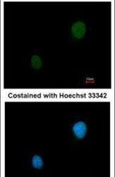
- Immunofluorescent analysis of PKA 2 beta in methanol-fixed HeLa cells using a PKA 2 beta polyclonal antibody (Product # PA5-28266) at a 1:200 dilution.
Supportive validation
- Submitted by
- Invitrogen Antibodies (provider)
- Main image
- Experimental details
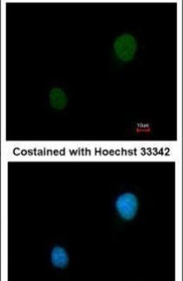
- Immunohistochemistry (Frozen) analysis of PRKAR2B was performed in frozen sectioned adult mouse hippocampus using PRKAR2B Polyclonal Antibody (Product # PA5-28266) at a dilution of 1:250. Blue: Fluoroshield with DAPI. Antigen Retrieval: Citrate buffer, pH 6.0, 10 min.

- Submitted by
- Invitrogen Antibodies (provider)
- Main image
- Experimental details
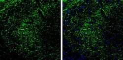
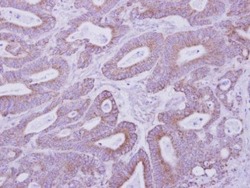
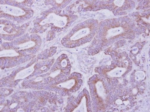
- Immunohistochemical analysis of paraffin-embedded human colon carcinoma, using PKA 2 beta (Product # PA5-28266) antibody at 1:500 dilution. Antigen Retrieval: Citrate buffer, pH 6.0, 10 min.
Supportive validation
- Submitted by
- Invitrogen Antibodies (provider)
- Main image

- Experimental details
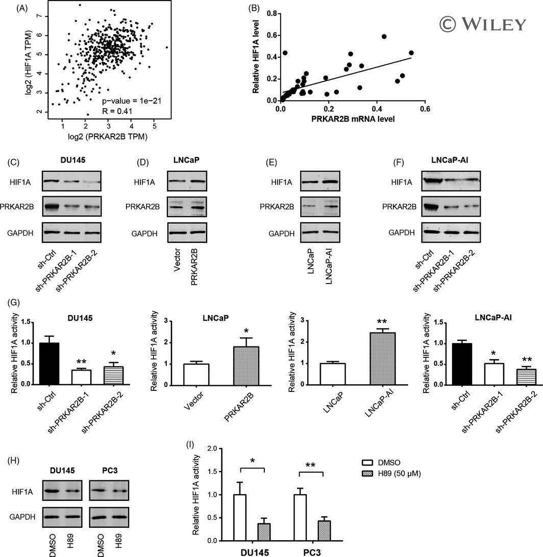
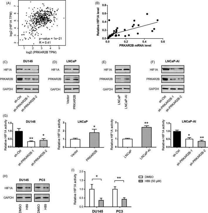
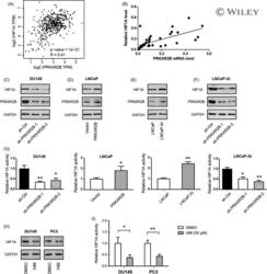
- FIGURE 4 PRKAR2B regulates HIF1alpha expression in prostate cancer. (A-B) Correlation analysis of the link between PRKAR2B and HIF1A expression in prostate cancer tissues; data were obtained from the TCGA cohort (A) and Ren Ji cohort (B). (C) Western blotting analysis of the effect of PRKAR2B knockdown on the HIF1A protein level in DU145 cells. (D) Western blotting analysis of the effect of PRKAR2B overexpression on the HIF1A protein level in LNCaP cells. (E) Western blotting analysis of HIF1A protein level in LNCaP and LNCaP-AI cells. (F) Western blotting analysis of the effect of PRKAR2B knockdown on the HIF1A protein level in LNCaP-AI cells. (G) Effects of PRKAR2B knockdown or overexpression on the HIF-1alpha activity. (H) Western blotting analysis of HIF1A protein level in DU145 and PC3 cells upon H89 (50 mumol/L) treatment for 24 h. (I) Determining the effect of H89 (50 mumol/L) treatment on the HIF-1alpha activity in DU145 and PC3 cells. * P < .05; ** P < .01
- Submitted by
- Invitrogen Antibodies (provider)
- Main image
- Experimental details
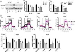
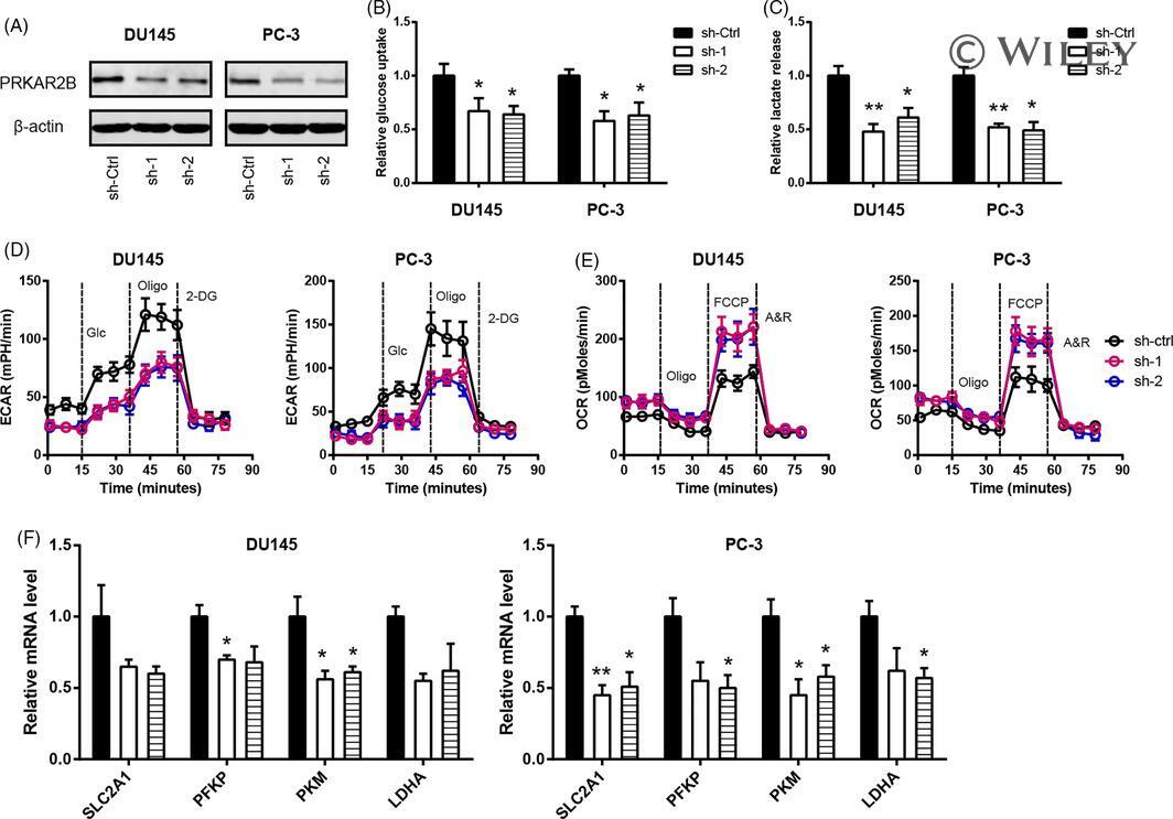
- 1 FIGURE PRKAR2B knockdown inhibits prostate cancer cell glycolysis. A, Western blotting analysis of the knockdown efficiency of PRKAR2B in DU145 and PC3 cells. B-E, The effect of PRKAR2B knockdown on the glucose utilization (B), lactate production (C), extracellular acidification rate (D), oxygen consumption rate (E), and expression of glycolytic components (F) in DU145 and PC3 cells. * P < .05; ** P < .01
- Submitted by
- Invitrogen Antibodies (provider)
- Main image

- Experimental details
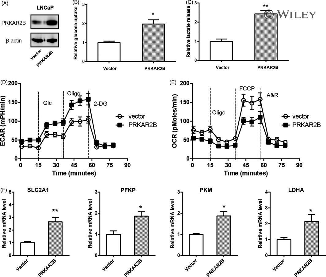
- 2 FIGURE PRKAR2B overexpression promotes the Warburg effect in prostate cancer in vitro. A, Western blotting analysis of the overexpression efficiency of PRKAR2B in LNCaP cells. B-E, The effect of PRKAR2B overexpression on the glucose utilization (B), lactate production (C), extracellular acidification rate (D), oxygen consumption rate (E), and expression of glycolytic components (F) in LNCaP cells. SLC2A1, solute carrier family 2 member 1; PFKP, phosphofructokinase platelet; PKM, pyruvate kinase muscle isozyme; LDHA, lactate dehydrogenase A. * P < .05; ** P < .01
- Submitted by
- Invitrogen Antibodies (provider)
- Main image
- Experimental details
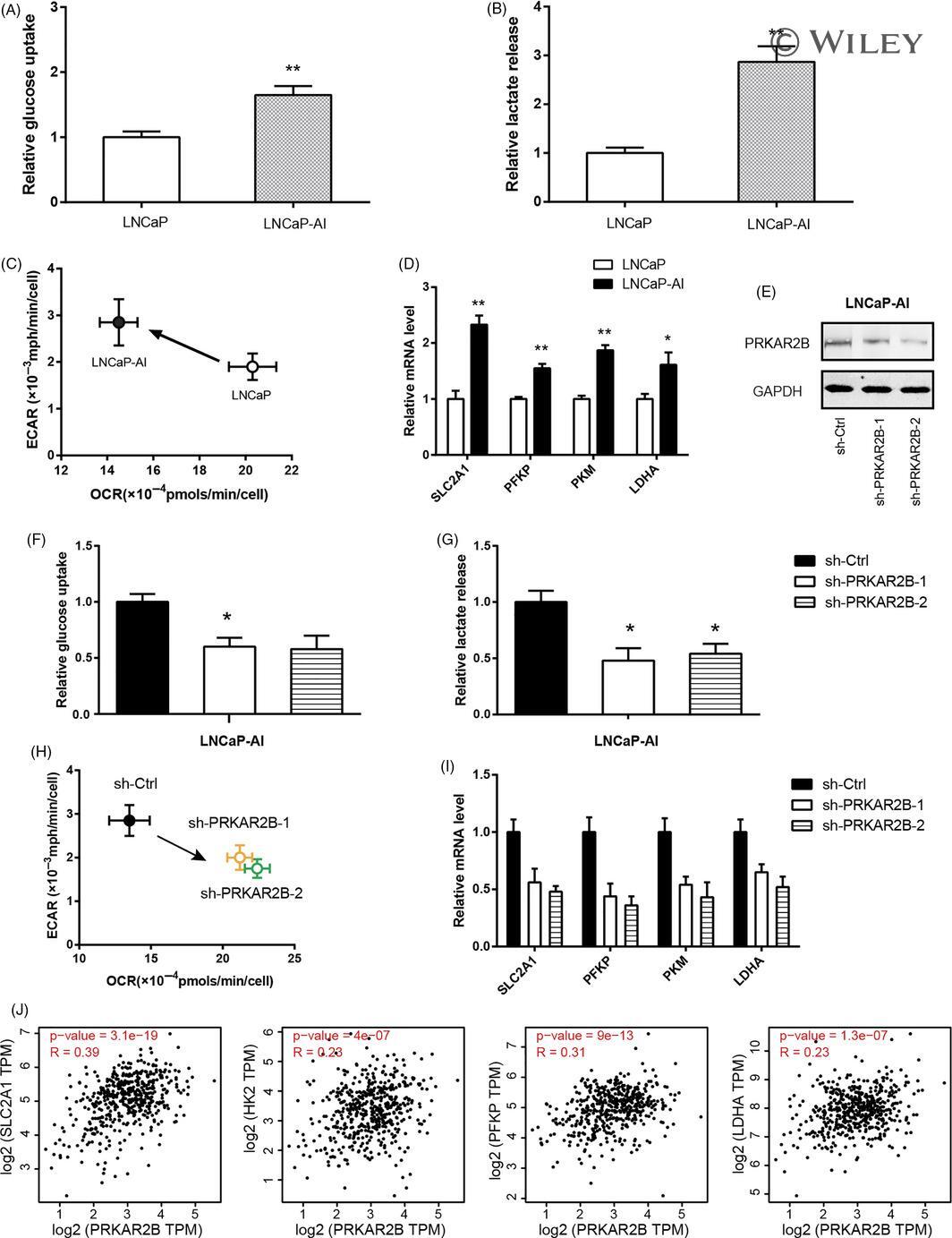
- 3 FIGURE Androgen-independent LNCaP cells exhibits enhanced glycolysis. A, The basal glucose utilization in LNCaP and LNCaP-AI cells. B, The basal lactate production in LNCaP and LNCaP-AI cells. C, Comparison of ECAR and OCR status in LNCaP and LNCaP-AI cells. D, Real-time qPCR analysis of glucose transporter and key glycolytic genes in LNCaP and LNCaP-AI cells. E, Western blotting analysis of the effect of PRKAR2B knockdown efficiency in LNCaP-AI cells. (F-I) The effect of PRKAR2B knockdown on the glucose utilization (F), lactate production (G), ECAR/OCR (H), and expression of glycolytic components (I) in LNCaP-AI cells. (J) Correlation analysis of the link between PRKAR2B expression and the expression level of glucose transporter and key glycolytic genes in prostate cancer tissues (n = 492); data were obtained from the TCGA cohort. * P < .05; ** P < .01
- Submitted by
- Invitrogen Antibodies (provider)
- Main image
- Experimental details
- 4 FIGURE PRKAR2B regulates HIF1alpha expression in prostate cancer. (A-B) Correlation analysis of the link between PRKAR2B and HIF1A expression in prostate cancer tissues; data were obtained from the TCGA cohort (A) and Ren Ji cohort (B). (C) Western blotting analysis of the effect of PRKAR2B knockdown on the HIF1A protein level in DU145 cells. (D) Western blotting analysis of the effect of PRKAR2B overexpression on the HIF1A protein level in LNCaP cells. (E) Western blotting analysis of HIF1A protein level in LNCaP and LNCaP-AI cells. (F) Western blotting analysis of the effect of PRKAR2B knockdown on the HIF1A protein level in LNCaP-AI cells. (G) Effects of PRKAR2B knockdown or overexpression on the HIF-1alpha activity. (H) Western blotting analysis of HIF1A protein level in DU145 and PC3 cells upon H89 (50 mumol/L) treatment for 24 h. (I) Determining the effect of H89 (50 mumol/L) treatment on the HIF-1alpha activity in DU145 and PC3 cells. * P < .05; ** P < .01