 Explore
 Validate
 Learn
Learn Western blot
Antibody data
- Antibody Data
- Antigen structure
- References [1]
- Comments [0]
- Validations
- Western blot [3]
- Immunocytochemistry [2]
- Immunoprecipitation [1]
- Other assay [1]
Submit
Validation data
Reference
Comment
Report error
- Product number
- PA5-65539 - Provider product page

- Provider
- Invitrogen Antibodies
- Product name
- ITCH Polyclonal Antibody
- Antibody type
- Polyclonal
- Antigen
- Recombinant protein fragment
- Description
- Immunogen sequence: GFTGASQNDDG SRSKDETRVS TNGSDDPEDA GAGENRRVSG NNSPSLSNGG FKPSRPPRPS RPPPPTPRRP ASVNGS Highest antigen sequence identity to the following orthologs - mouse 78%, rat 82%.
- Reactivity
- Human
- Host
- Rabbit
- Isotype
- IgG
- Vial size
- 100 μL
- Concentration
- 0.2 mg/mL
- Storage
- Store at 4°C short term. For long term storage, store at -20°C, avoiding freeze/thaw cycles.
Submitted references IL-33 enhances Jagged1 mediated NOTCH1 intracellular domain (NICD) deubiquitination and pathological angiogenesis in proliferative retinopathy.
Sharma D, Bisen S, Kaur G, Van Buren EC, Rao GN, Singh NK
Communications biology 2022 May 19;5(1):479
Communications biology 2022 May 19;5(1):479
No comments: Submit comment
Supportive validation
- Submitted by
- Invitrogen Antibodies (provider)
- Main image

- Experimental details
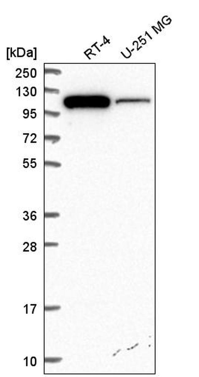
- Western blot analysis of ITCH in human cell line RT-4 and human cell line U-251 MG. Samples were probed using an ITCH Polyclonal Antibody (Product # PA5-65539).
- Submitted by
- Invitrogen Antibodies (provider)
- Main image
- Experimental details
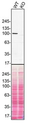
- Western blot of ITCH was performed by loading 100 µg of WT (lane 1) and ITCH CRISPR KO (lane 2) HeLa cell lysates in RIPA buffer onto a 4-15% gradient polyacrylamide gel. Proteins on the blots were visualized with Ponceau staining (below immunoblot). Proteins were transferred to nitrocellulose membrane and blocked in 5% milk for 1 hr. ITCH was detected at approximately 102 kDa using an ITCH polyclonal antibody (Product # PA5-65539) at a dilution of 1:1,000 in 5% BSA in TBS with 0.1% Tween 20 (TBST) overnight at 4°C. The peroxidase-conjugated secondary antibody (Product # 65-6120) was diluted to 0.2 µg/mL in TBST with 5% milk for 1 hr. Chemiluminescent detection was performed using Pierce ECL Western Blotting Substrate (Product # 32106). Data courtesy of YCharOS Inc., an open science company with the mission of characterizing commercially available antibodies using knockout validation.

- Submitted by
- Invitrogen Antibodies (provider)
- Main image
- Experimental details
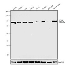
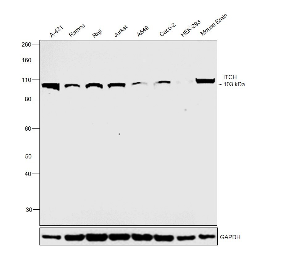
- Western blot was performed using Anti-ITCH Polyclonal Antibody (Product # PA5-65539) and a 103 kDa band corresponding to ITCH was observed across cell lines and tissue extracts tested. Whole cell extracts (30 µg lysate) of A-431 (Lane 1), Ramos (Lane 2), Raji (Lane 3), Jurkat (Lane 4), A549 (Lane 5), Caco-2 (Lane 6), HEK-293 (Lane 7) and tissue extract of Mouse Brain (Lane 8) were electrophoresed using NuPAGE™ 4-12% Bis-Tris Protein Gel (Product # NP0322BOX). Resolved proteins were then transferred onto a nitrocellulose membrane (Product # IB23001) by iBlot® 2 Dry Blotting System (Product # IB21001). The blot was probed with the primary antibody (1:500 dilution) and detected by chemiluminescence with Goat anti-Rabbit IgG (Heavy Chain) Superclonal™ Recombinant Secondary Antibody, HRP (Product # A27036, 1:4000 dilution) using the iBright FL 1000 (Product # A32752). Chemiluminescent detection was performed using Novex® ECL Chemiluminescent Substrate Reagent Kit (Product # WP20005).
Supportive validation
- Submitted by
- Invitrogen Antibodies (provider)
- Main image
- Experimental details
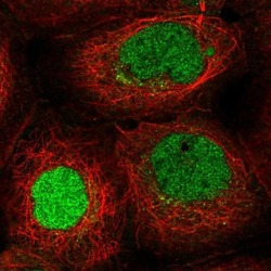
- Immunofluorescent staining of ITCH in human cell line A-431 shows localization to nucleoplasm. Samples were probed using an ITCH Polyclonal Antibody (Product # PA5-65539).

- Submitted by
- Invitrogen Antibodies (provider)
- Main image

- Experimental details
- Immunofluorescent staining of ITCH in human cell line A-431 shows localization to nucleoplasm. Samples were probed using an ITCH Polyclonal Antibody (Product # PA5-65539).

Supportive validation
- Submitted by
- Invitrogen Antibodies (provider)
- Main image
- Experimental details
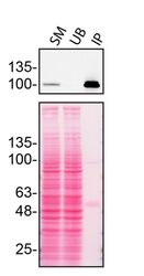
- Immunoprecipitation of ITCH was performed on HeLa cell lysates. Antibody-bead conjugates were prepared by adding 2 µg of ITCH polyclonal antibody (Product # PA5-65539) with 30 µL of protein G-Sepharose beads and rocked overnight at 4°C. 750 µg of lysate was incubated with an antibody-bead conjugate for 2 hours at 4°C. Following centrifugation and multiple washes, 10% starting material (SM), 10% unbound fraction (UB) and immunoprecipitated fraction (IP) were processed for immunoblot using a different antibody. Ponceau stained transfer of blot is shown. Data courtesy of YCharOS Inc., an open science company with the mission of characterizing commercially available antibodies using knockout validation.

Supportive validation
- Submitted by
- Invitrogen Antibodies (provider)
- Main image

- Experimental details
- Immunoprecipitation of ITCH was performed on HeLa cell lysates. Antibody-bead conjugates were prepared by adding 2 µg of ITCH polyclonal antibody (Product # PA5-65539) with 30 µL of protein G-Sepharose beads and rocked overnight at 4°C. 750 µg of lysate was incubated with an antibody-bead conjugate for 2 hours at 4°C. Following centrifugation and multiple washes, 10% starting material (SM), 10% unbound fraction (UB) and immunoprecipitated fraction (IP) were processed for immunoblot using a different antibody. Ponceau stained transfer of blot is shown. Data courtesy of YCharOS Inc., an open science company with the mission of characterizing commercially available antibodies using knockout validation.
