Explore
 Validate
 Learn
Learn Western blot
Antibody data
- Antibody Data
- Antigen structure
- References [4]
- Comments [0]
- Validations
- Western blot [2]
- Immunohistochemistry [1]
- Flow cytometry [3]
- Other assay [2]
Submit
Validation data
Reference
Comment
Report error
- Product number
- 700043 - Provider product page

- Provider
- Invitrogen Antibodies
- Product name
- Phospho-PKC theta (Thr538) Recombinant Rabbit Monoclonal Antibody (F4H4L1)
- Antibody type
- Monoclonal
- Antigen
- Synthetic peptide
- Description
- This antibody is predicted to react with bovine, chimpanzee, mouse, rat and Xenopus based on sequence homology. Intact IgG appears on a non-reducing gel as ~150 kDa band and upon reduction generating a ~25 kDa light chain band and a ~50 kDa heavy chain. Recombinant rabbit monoclonal antibodies are produced using in vitro expression systems. The expression systems are developed by cloning in the specific antibody DNA sequences from immunoreactive rabbits. Then, individual clones are screened to select the best candidates for production. The advantages of using recombinant rabbit monoclonal antibodies include: better specificity and sensitivity, lot-to-lot consistency, animal origin-free formulations, and broader immunoreactivity to diverse targets due to larger rabbit immune repertoire.
- Reactivity
- Human
- Host
- Rabbit
- Isotype
- IgG
- Antibody clone number
- F4H4L1
- Vial size
- 100 μg
- Concentration
- 0.5 mg/mL
- Storage
- Store at 4°C short term. For long term storage, store at -20°C, avoiding freeze/thaw cycles.
Submitted references PD-1 is a haploinsufficient suppressor of T cell lymphomagenesis.
Intracellular Delivery of Anti-pPKCθ (Thr538) via Protein Transduction Domain Mimics for Immunomodulation.
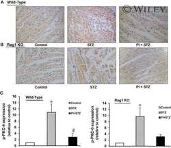
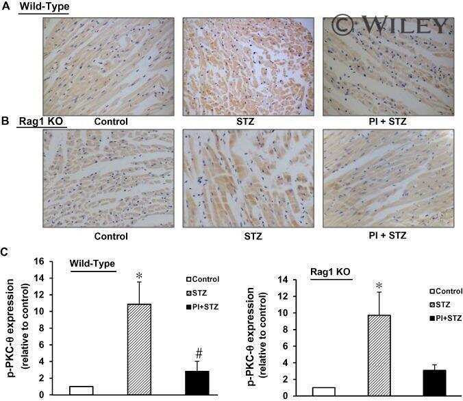
Inhibition of PKC-θ preserves cardiac function and reduces fibrosis in streptozotocin-induced diabetic cardiomyopathy.
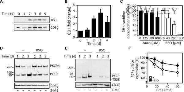
PKC-θ exists in an oxidized inactive form in naive human T cells.
Wartewig T, Kurgyis Z, Keppler S, Pechloff K, Hameister E, Öllinger R, Maresch R, Buch T, Steiger K, Winter C, Rad R, Ruland J
Nature 2017 Dec 7;552(7683):121-125
Nature 2017 Dec 7;552(7683):121-125
Intracellular Delivery of Anti-pPKCθ (Thr538) via Protein Transduction Domain Mimics for Immunomodulation.
Ozay EI, Gonzalez-Perez G, Torres JA, Vijayaraghavan J, Lawlor R, Sherman HL, Garrigan DT Jr, Burnside AS, Osborne BA, Tew GN, Minter LM
Molecular therapy : the journal of the American Society of Gene Therapy 2016 Dec;24(12):2118-2130
Molecular therapy : the journal of the American Society of Gene Therapy 2016 Dec;24(12):2118-2130
Inhibition of PKC-θ preserves cardiac function and reduces fibrosis in streptozotocin-induced diabetic cardiomyopathy.
Li Z, Abdullah CS, Jin ZQ
British journal of pharmacology 2014 Jun;171(11):2913-24
British journal of pharmacology 2014 Jun;171(11):2913-24
PKC-θ exists in an oxidized inactive form in naive human T cells.
von Essen MR, Kongsbak M, Levring TB, Hansen AK, Boding L, Lauritsen JP, Woetmann A, Baier G, Ødum N, Bonefeld CM, Geisler C
European journal of immunology 2013 Jun;43(6):1659-66
European journal of immunology 2013 Jun;43(6):1659-66
No comments: Submit comment
Supportive validation
- Submitted by
- Invitrogen Antibodies (provider)
- Main image

- Experimental details
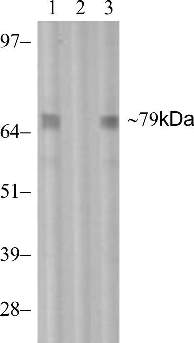
- Western blot analysis of Phospho-PKC theta pThr538 in Jurkat cell lysate stimulated with 100 ng/mL PMA for 1 hr using a Phospho-PKC theta pThr538 recombinant rabbit monoclonal antibody (Product # 700043) at a dilution of 3 µg/mL.
- Submitted by
- Invitrogen Antibodies (provider)
- Main image
- Experimental details
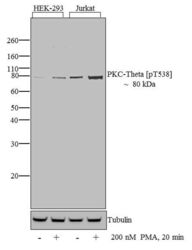
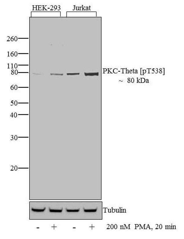
- Western blot analysis of PKC-theta (pT538) was performed by loading 30 µg of HEK-293 (lane1) HEK-293 treated for 20 minutes with 200 nM of PMA (lane2), Jurkat (lane3) and Jurkat treated for 20 minutes with 200 nM of PMA (lane4) cell lysate using Novex®NuPAGE® 12 % Bis-Tris gel (Product # NP0342BOX), XCell SureLock Electrophoresis System (Product # EI0002), Novex® Sharp Pre-Stained Protein Standard (LC5800), and iBlot® 2 Dry Blotting System (IB21001). Proteins were transferred to a nitrocellulose membrane and blocked with 5% skim milk for 1 hour at room temperature. PKC-theta (pT538) was detected at ~80 kDa using PKC-theta (pT538) Rabbit Monoclonal Antibody (Product # 700043) at 1-2 µg/mL in 5% skim milk at 4°C overnight on a rocking platform. Goat Anti-Rabbit IgG - HRP Secondary Antibody (G21234) at 1:5000 dilution was used and chemiluminescent detection was performed using Pierce™ ECL Western Blotting Substrate (Product # 32106).
Supportive validation
- Submitted by
- Invitrogen Antibodies (provider)
- Main image

- Experimental details
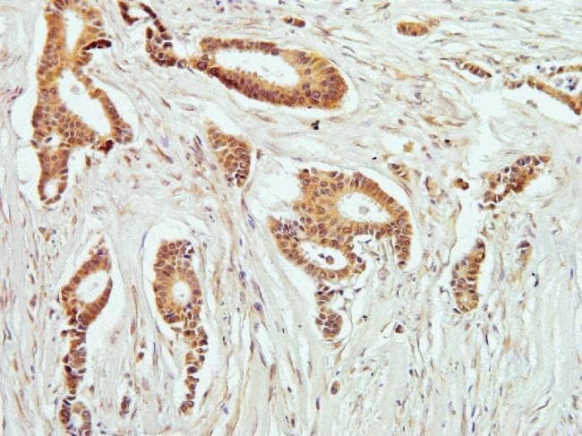
- Immunohistochemistry analysis of Phospho-PKC theta pThr538 in formalin-fixed, paraffin-embedded human breast carcimona tissue using a Phospho-PKC theta pThr538 monoclonal antibody (Product # 700043) at a dilution of 5 µg/mL. Staining was visualized using DAB. Images were taken at a magnification of 20x. Results show cytoplasmic staining in tumor cells.
Supportive validation
- Submitted by
- Invitrogen Antibodies (provider)
- Main image
- Experimental details
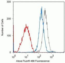
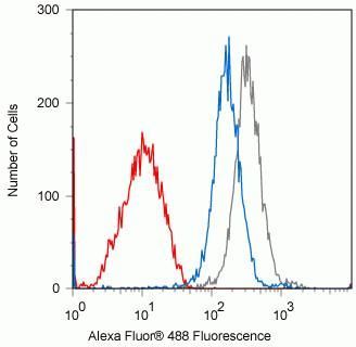
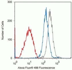
- Flow cytometry analysis of Phospho-PKC theta pThr538 in Jurkat cells treated with 100uM PMA for 1hr using a Phospho-PKC theta pThr538 recombinant rabbit monoclonal antibody (Product # 700043) at a dilution of 0.1 µg. Cells were fixed and permeabilized using FIX & PERM (Product # GAS-004) reagent, and detection was performed using an Alexa Fluor 488 goat anti-rabbit IgG (gray) compared to samples incubated with a non-phosphopeptide (blue). Pre-incubation with the immunogenic peptide decreased the signal (red) whereas incubation with the non-phosphopeptide did not.
- Submitted by
- Invitrogen Antibodies (provider)
- Main image
- Experimental details
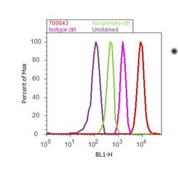
- Flow cytometry analysis of PKC-theta [pT538] was done on Jurkat cells treated with PMA (200nM, 20 minutes). Cells were fixed with 70% ethanol for 10 minutes, permeabilized with 0.25% Tritonª X-100 for 20 minutes, and blocked with 5% BSA for 30 minutes at room temperature. Cells were labeled with ABfinityª PKC-theta [pT538] Recombinant Rabbit Monoclonal Antibody (700043, red histogram) or with rabbit isotype control (pink histogram) at 3-5 µg/million cells in 2.5% BSA. After incubation at room temperature for 2 hours, the cells were labeled with Alexa Fluor¨ 488 Goat Anti-Rabbit Secondary Antibody (A11008) at a dilution of 1:400 for 30 minutes at room temperature. The representative 10,000 cells were acquired and analyzed for each sample using an Attune¨ Acoustic Focusing Cytometer. The purple histogram represents unstained control cells and the green histogram represents no-primary-antibody control.

- Submitted by
- Invitrogen Antibodies (provider)
- Main image
- Experimental details
- Flow cytometry analysis of Phospho-PKC theta pThr538 in Jurkat cells treated with 100uM PMA for 1hr using a Phospho-PKC theta pThr538 recombinant rabbit monoclonal antibody (Product # 700043) at a dilution of 0.1 µg. Cells were fixed and permeabilized using FIX & PERM (Product # GAS-004) reagent, and detection was performed using an Alexa Fluor 488 goat anti-rabbit IgG (gray) compared to samples incubated with a non-phosphopeptide (blue). Pre-incubation with the immunogenic peptide decreased the signal (red) whereas incubation with the non-phosphopeptide did not.

Supportive validation
- Submitted by
- Invitrogen Antibodies (provider)
- Main image
- Experimental details
- NULL
- Submitted by
- Invitrogen Antibodies (provider)
- Main image

- Experimental details
- NULL