 Explore
 Validate
 Learn
Learn Western blot
Antibody data
- Antibody Data
- Antigen structure
- References [1]
- Comments [0]
- Validations
- Western blot [1]
- Immunocytochemistry [1]
Submit
Validation data
Reference
Comment
Report error
- Product number
- 14-0498-37 - Provider product page

- Provider
- Invitrogen Antibodies
- Product name
- CD49b (Integrin alpha 2) Monoclonal Antibody (eBioY418 (Y418)), eBioscience™
- Antibody type
- Monoclonal
- Antigen
- Other
- Description
- Description: The eBioY418 (Y418) monoclonal antibody reacts with human CD49b, the 150-kDa, integrin alpha 2 subunit. The complex of CD49b non-covalently associated with integrin beta 1 (CD29), also known as VLA-2, is a receptor for collagen and laminin. This complex is expressed by lymphocytes, activated T cells, monocytes and platelets.
- Antibody clone number
- eBioY418 (Y418)
- Concentration
- 0.5 mg/mL
Submitted references Prevailing role of contact guidance in intrastromal T-cell trapping in human pancreatic cancer.
Hartmann N, Giese NA, Giese T, Poschke I, Offringa R, Werner J, Ryschich E
Clinical cancer research : an official journal of the American Association for Cancer Research 2014 Jul 1;20(13):3422-33
Clinical cancer research : an official journal of the American Association for Cancer Research 2014 Jul 1;20(13):3422-33
No comments: Submit comment
Supportive validation
- Submitted by
- Invitrogen Antibodies (provider)
- Main image
- Experimental details
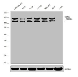
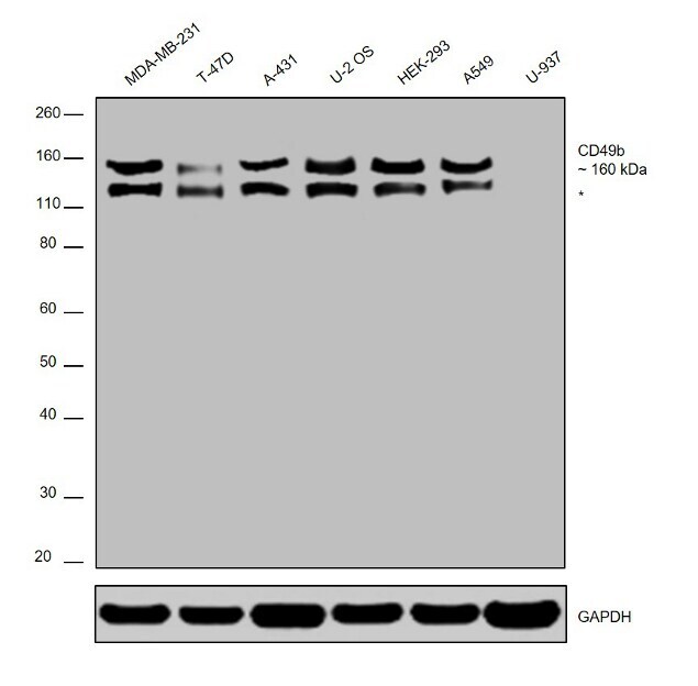
- Western blot was performed using Anti-CD49b Polyclonal Antibody (Product # 14-0498-80) and a 160kDa band corresponding to CD49b was observed across cell lines tested except U-937. Whole cell extracts (30 µg lysate) of MDA-MB-231 (Lane 1), T-47D (Lane 2), A-431 (Lane 3), U-2 OS (Lane 4), HEK-293 (Lane 5), A549 (Lane 6) and U-937 (Lane 7) were electrophoresed using NuPAGE™ 4-12% Bis-Tris Protein Gel (Product # NP0322BOX). Resolved proteins were then transferred onto a nitrocellulose membrane (Product # IB23001) by iBlot® 2 Dry Blotting System (Product # IB21001). The blot was probed with the primary antibody (0.25 µg/mL) and detected by chemiluminescence Goat anti-Rabbit IgG (H+L) Superclonal™ Secondary Antibody, HRP (Product # A27036, 1:4000 dilution) using the iBright FL 1000 (Product # A32752). Chemiluminescent detection was performed using Novex® ECL Chemiluminescent Substrate Reagent Kit (Product # WP20005). Note: An uncharacterized band (*) at ~125 kDa was observed in all the samples except U937.
Supportive validation
- Submitted by
- Invitrogen Antibodies (provider)
- Main image
- Experimental details
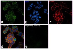
- Immunofluorescence analysis of CD49b/ITGA2 was performed using 70% confluent log phase U-2 OS cells. The cells were fixed with 4% paraformaldehyde for 10 minutes and blocked with 2% BSA for 1 hour at room temperature. The cells were labeled with CD49b/ITGA2 Mouse Monoclonal Antibody (Product # 14-0498-80) at 5 µg/mL in 0.1% BSA, incubated at 4 degree Celsius overnight and then labeled with Goat anti-Mouse IgG (H+L) Superclonal™ Secondary Antibody, Alexa Fluor® 488 conjugate (Product # A28175) at a dilution of 1:2000 for 45 minutes at room temperature (Panel a: green). Nuclei (Panel b: blue) were stained with ProLong™ Diamond Antifade Mountant with DAPI (Product # P36962). F-actin (Panel c: red) was stained with Rhodamine Phalloidin (Product # R415). Panel d represents the merged image showing membrane localization. Panel e represents control cells with no primary antibody to assess background. The images were captured at 60X magnification.
