 Explore
 Validate
 Learn
Learn Western blot
Antibody data
- Antibody Data
- Antigen structure
- References [1]
- Comments [0]
- Validations
- Western blot [2]
- Other assay [1]
Submit
Validation data
Reference
Comment
Report error
- Product number
- PA5-26061 - Provider product page

- Provider
- Invitrogen Antibodies
- Product name
- ITGA2 Polyclonal Antibody
- Antibody type
- Polyclonal
- Antigen
- Synthetic peptide
- Reactivity
- Human
- Host
- Rabbit
- Isotype
- IgG
- Vial size
- 400 µL
- Concentration
- 0.5 mg/mL
- Storage
- Store at 4°C short term. For long term storage, store at -20°C, avoiding freeze/thaw cycles.
Submitted references ITGA2 promotes expression of ACLY and CCND1 in enhancing breast cancer stemness and metastasis.
Adorno-Cruz V, Hoffmann AD, Liu X, Dashzeveg NK, Taftaf R, Wray B, Keri RA, Liu H
Genes & diseases 2021 Jul;8(4):493-508
Genes & diseases 2021 Jul;8(4):493-508
No comments: Submit comment
Supportive validation
- Submitted by
- Invitrogen Antibodies (provider)
- Main image

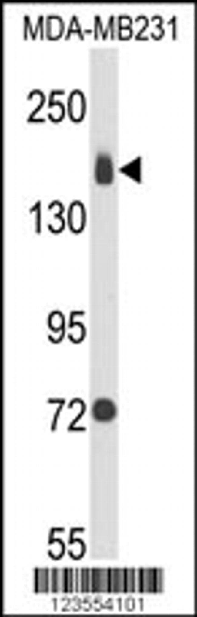
- Experimental details
- Western blot analysis using an ITGA2 polyclonal antibody (Product # PA5-26061) in MDA-MB231 cell lysates (35 µg per lane).

- Submitted by
- Invitrogen Antibodies (provider)
- Main image

- Experimental details
- Western blot analysis of ITGA2 in A431 whole cell lysates (20 µg/lane). Samples were probed with an ITGA2 Antibody (C-term) (Product # PA5-26062) at 1:1000 dilution, followed by incubation with a Secondary Goat Anti-Rabbit IgG, (H+L), Peroxidase conjugated at 1:10000 dilution. Predicted band size : 129 kDa. Blocking/Dilution buffer: 5% NFDM/TBST.
Supportive validation
- Submitted by
- Invitrogen Antibodies (provider)
- Main image

- Experimental details
- Figure 2 ITGA2 is a target of miR-206 and promotes mammosphere formation and stemness factor expression. ( A-B ) ITGA2 mRNA (top panel) and protein (bottom panel) levels are reduced by transfected miR-206 in MDA-MB-231 cells, measured by qRT-PCR and immunoblotting, respectively. *** t- test P < 0.001. Error bars represent S.D. values. ( C ) CD49b surface protein expression inhibited by miR-206 in MDA-MB-231 cells, evaluated by flow cytometry. ( D ) Predicted binding sites between the complementary sequences of ITGA2 3'UTR (WT) and miR-206. The bottom line shows the mutated C to T (U, in red) within the interaction site of 3'UTR. ( E ) 3'UTR luciferase assay shows direct regulation of ITGA2 by miR-206, n = 3, ** t- test P = 0.005. ( F-G ) Images (F) and quantified counts (G) of breast cancer cell-derived mammospheres upon si ITGA2 knockdown. ** t- test P < 0.01. Error bars represent S.D. values. Scale bars = 25 mum ( H-I ) siITGA2 transfection depletes CD49b protein expression, measured by immunoblotting (H) and flow cytometry analyses (I). ( J ) Immunoblots to detect reduced expression of CD49b, phosphorylated FAK (pFAK), and OCT3/4 levels without affecting total FAK levels upon ITGA2 KD or miR-206 upregulation. ( K ) Left panel: Flow histogram of CD49b expression in control and siITGA2-transfected cells and gated sorting of CD49b ++ and CD49b low/- populations, respectively. Right panel: Bioluminescent images of CD49b ++ and CD49b low/- implants of 100-1000 cells from day 1-
